Figures & data
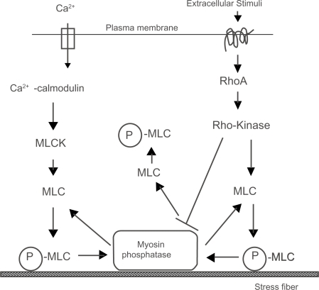
Figure 1 The localization of stress fibers (a and d) and focal adhesion (c and d) in cultured endothelial cells. The cells were stained with rhodamine-labeled phalloidin for F-actin (stress fiber) visualization (a and b) or antivinculin for focal adhesion visualization (c and d). Micrographs are taken at two focal planes: a and c are adjusted at the apical portion of the cell; b and d are adjusted at the basal portion of the cell. Apical stress fibers are clearly visible (a; arrows) together with the typical basal stress fibers (b). Many of the anti-vinculin stained spots are observed at the apical portion of the cell (c; arrows) together with the staining of basal focal adhesions (d).
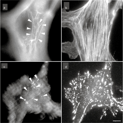
Figure 2 Confocal laser scanning optical images of in situ endothelial cells in guinea pig aorta endothelial cells were doubly stained with rhodamine-labeled phalloidin for F-actin (stress fiber) visualization (a, c, and e) and with propidium iodide for nuclei (b, d, and f). Stress fibers are observed at both the apical side of the cell (arrows) and the basal side of the cell (b), along with the direction of the blood flow (b; flow). e and f; A side view of the endothelium. Apical stress fibers (e; arrows) and the F-actin accumulation of cell-cell apposition sites (e; arrows) are visible.
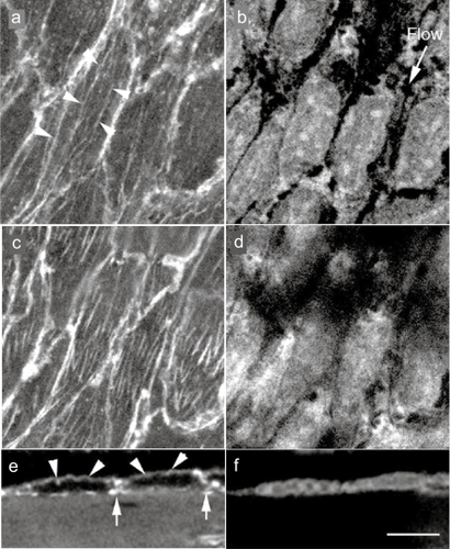
Figure 3 Stress fibers in in situ endothelial cells in a low blood flow area (a) and a high blood flow area (b). In the low blood flow area, cell shape is circular and the expression of stress fibers is low (a). In the direction of high blood flow, the endothelial cells are elongated and the expression of stress fibers is increased along the direction of the blood flow (b).
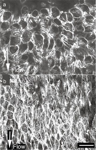
Figure 4 Schematic illustrations of the stress fibers, focal adhesions, and apical plaques in endothelial cells. Stress fibers run not only on the basal side of the cell, but they also run from the basal side to the apical portion of the cell. Focal adhesion-like structures, called “apical plaques” are localized at the apical side of the endothelial cells in situ.
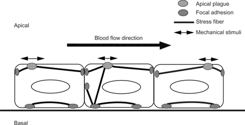
Figure 5 A schematic illustration of two regulatory systems for actomyosin contraction in nonmuscle cells. Phosphorylated MLC (P-MLC) induces contraction and the organization of stress fibers.