Figures & data
Table 1 Gene-specific primer sequences and accession numbers
Figure 1 The expression of the inflammatory genes tended to be greater in the cerebral arteries treated with DSP than in the cerebral arteries treated with DMSO control. There was a significant upregulation of the extracellular-matrix-related genes (MMP9 and MMP13) in the DSP group, compared with DMSO control. The cerebral arteries have been organ cultured for 24 hours in the presence of DSP or DMSO (control).
Abbreviations: DMSO, dimethyl sulfoxide; DSP, cigarette smoke particles; MMP, matrix metalloproteinase; ns, not significant.
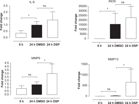
Figure 2 The expression of angiotensin II receptors in the cerebral arteries following organ culture with DSP or DMSO (control), there was a significant upregulation of the AT1 receptors induced by DSP compared with DMSO.
Abbreviations: AT, angiotensin receptor; DMSO, dimethyl sulfoxide; DSP, cigarette smoke particles; ns, not significant.
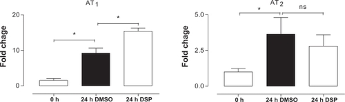
Figure 3 No p38 phosphorylation was found in the fresh cerebral arteries, while a slight increase was obtained in the cerebral arteries treated with DMSO and a strong activation in the cerebral arteries treated with DSP, ATF-2 and Elk-1 showed the same pattern as the p38 phosphorylation. However, phosphorylation of Elk-1 was also seen in the fresh cerebral arteries.
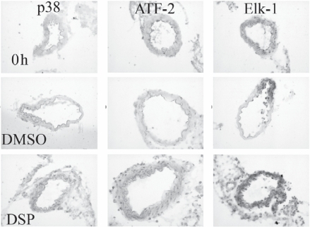
Table 2 Phosphorylation of intracellular signal proteins induced by DSP
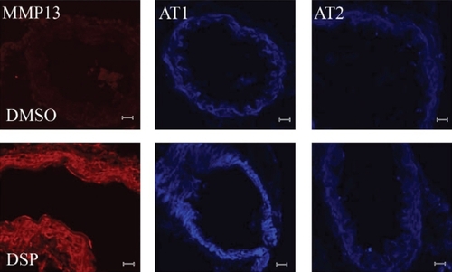
Figure 4 Immunohistochemistry shows a significant increase in the amount of phosphorylated MMP13 proteins and AT1 receptor proteins in the cerebral arteries treated with DSP, compared with the control (DMSO-treated cerebral arteries). However, there was no increase in AT2 receptor proteins induced by DSP.
Abbreviations: AT, angiotensin receptor; DMSO, dimethyl sulfoxide; DSP, cigarette smoke particles; MMP, matrix metalloproteinase.