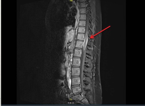
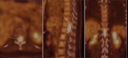
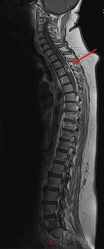
1,572
Views
0
CrossRef citations to date
0
Altmetric
Case Report

Extranodal classical Hodgkin lymphoma involving the spinal cord: case report and review of the literature

ORCID Icon, , , , , , , , , & show all
Article: CNS88 | Received 26 Jun 2021, Accepted 26 Apr 2022, Published online: 13 Jun 2022

Figures & data

Figure 1. Patient 1.

Sagittal T1-weighted MRI showing an intra medullary spinal cord lesion from T10 to L1 levels (arrow).

Figure 1. Patient 1. Sagittal T1-weighted MRI showing an intra medullary spinal cord lesion from T10 to L1 levels (arrow).
Figure 2. Atypical spinal cord lymphoma infiltration.

Transversal and sagittal slides from PET showing intramedullary signal in front of T10 to L1 vertebrae. The arrows show a hypermetabolic lesion located in the spinal cord.

Figure 2. Atypical spinal cord lymphoma infiltration. Transversal and sagittal slides from PET showing intramedullary signal in front of T10 to L1 vertebrae. The arrows show a hypermetabolic lesion located in the spinal cord.
Figure 3. Patient 2.

Sagittal T1-weighted MRI showing an intramedullary mass regarding T2 vertebrae (arrow).

Figure 3. Patient 2. Sagittal T1-weighted MRI showing an intramedullary mass regarding T2 vertebrae (arrow).

Table 1. Literature review.