Figures & data
Side-by-side comparison of post contrast, T1-weighted MRIs done before (left-sided panels) and 6 weeks after initiation of T-DXd. Each arrow indicates a metastasis that resolved after treatment initiation.
TDXd: Trastuzumab deruxtecan.
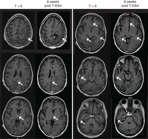
Table 1. Metastasis evolution after 2 months of initial (1A) and subsequent (1B) T-DXd administration.
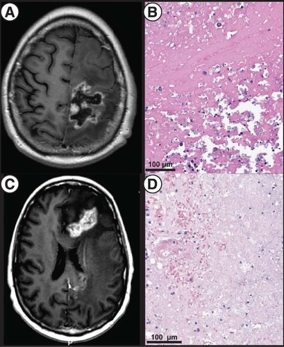
(A) T1-weighted, post contrast MRI showing prominent size of a dominant left frontoparietal lesion. (B) Postoperative pathology showing necrotic and fibrinous material and, at upper right, dystrophic mineralization without evidence of active tumor (H&E staining). (C) T1-weighted, post contrast MRI showing a dominant, bilobed left frontal lesion with surrounding vasogenic edema responsible for the patient’s symptoms and treated via LITT. (D) Pathology of the targeted lesion showing tissue necrosis with few mononuclear cells (H&E).
LITT: Laser-interstitial thermal therapy.
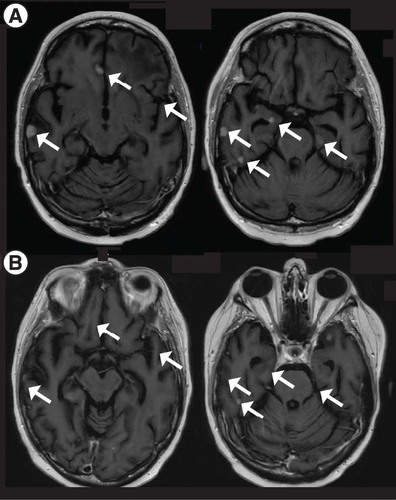
Post contrast T1-weighted MRI showing (A) progression of CNS disease in the setting of a T-DXd holiday and (B) improvement of most lesion upon T-DXd reinstatement (2 months after (A)). Each arrow indicates a metastasis that resolved after treatment initiation.
TDXd: Trastuzumab deruxtecan.