Figures & data
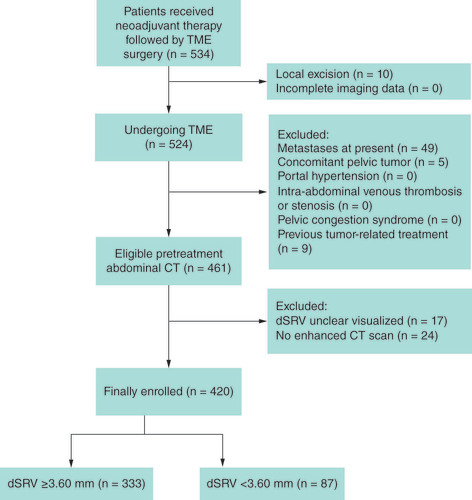
dSRV: Diameter of the superior rectal vein; TME: Total mesorectal excision.
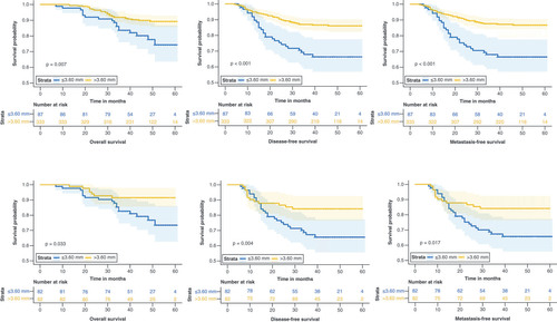
dSRV: Diameter of the superior rectal vein.
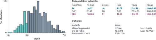
Table 1. Comparison of patient characteristics according to diameter of the superior rectal vein.
The first row shows the survival curve prior to propensity score matching.
SRV: Superior rectal vein.