Figures & data
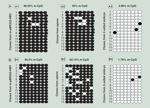
(A) Detection of CpG site methylation in island II. (A1) in plasmid; (A2) in sperm; and (A3) in sperm-derived embryo. (B) Detection of CpG site methylation in island III. (B1) in plasmid; (B2) in sperm; and (B3) in sperm-derived embryo. The methylation statuses of 14 clones for each sample are presented; each column represents one CpG position in island II or III, and each circle in the column indicates either cytosine (open circles) or methyl cytosine (filled circles).
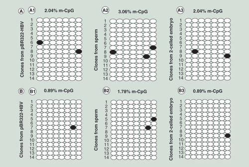
(A) Detection of CpG site methylation in island II. (A1) in plasmid; (A2) in sperm; and (A3) in sperm-derived embryo. (B) Detection of CpG site methylation in island III. (B1) in plasmid; (B2) in sperm; and (B3) in sperm-derived embryos.
(A) In sperm. M: marker DL2000; 1: X gene; 2: S gene; 3: -RT; 4: positive control; 5: β-actin; and 6: -T. (B) In sperm-derived embryos. M: marker DL2000; 1: X gene; 2: S gene; 3: -T; 4: -RT; 5: positive control; 6: β-actin.
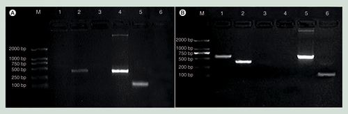
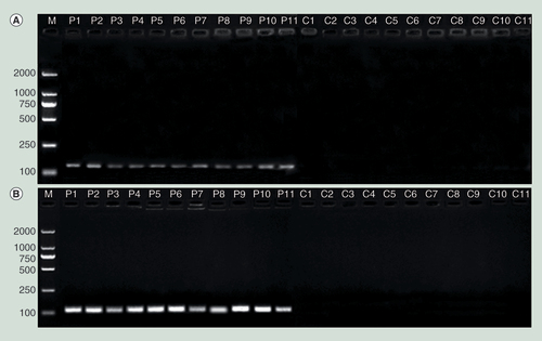
Methylation-specific bands for CpG sites in island II (A) and island III (B) in the hepatitis B virus genome were observed for the patient sperm but not for the control sperm.
C1–C11: Sperm from controls; M: Marker; P1–P11: Sperm from the patients.
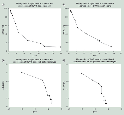
The transcriptional levels of the X and S genes increased with decreases in the methylation percentages of CpG sites in islands II and III, respectively, and strong negative correlations were observed. (A) X gene/island II correlation in sperm (Spearman’s r = -1; p < 0.0001); (B) X gene/island II correlation in sperm-derived embryos (Spearman’s r = -0.9623; p < 0.0001); (C) S gene/island III correlation in sperm (Spearman’s r = -0.9909; p < 0.0001); and (D) S gene/island III correlation in sperm-derived embryos (Spearman’s r = -0.9745; p < 0.0001).
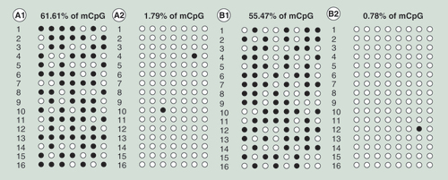
(A1) Island II with S-adenosylmethionine (SAM); (A2) island II without SAM; (B1) island III with SAM; and (B2) island III without SAM. The results showed that the methylation percentages of CpG sites in islands II and III in the embryos markedly increased following treatment with SAM.