Figures & data
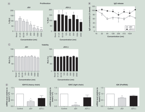
(A) JQ1 but not JQ1(-) significantly reduced cell proliferation, measured by the BrdU assay, at concentrations of 250 nM or higher (n = 6), data shown as percentage relative light units (RLU), relative to the untreated control. (B) IgG release, measured by an agglutination assay, was significantly reduced by treatment with JQ1 but not JQ1(-) (n = 5). (C) Cell viability, measured by the MTT assay, was not reduced by treatment of the CLNH11.4 cells with either JQ1 or JQ1(-) at any of the concentrations used during this study (n = 7). (D) Immunoglobulin encoding mRNA expressions from both the heavy (IGH1G) and light (IGKC) chains were significantly reduced by treatment with 200 μM of JQ1 but not JQ1(-), as was the preRNA from the κ light chain (n = 3). All data are shown as the mean ± the standard error.
*p < 0.05; **p < 0.01; ***p < 0.001 relative to control.
RLU: Relative light unit.
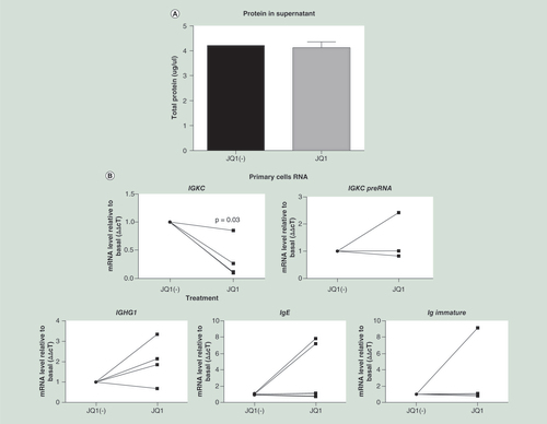
Cells were isolated and grown in culture for 7 days, after which they were treated with 500 nM JQ1 or JQ1(-) for a further 3 days. Immunoglobulin production was measured as (A) protein in the supernatant, and (B) gene expression including light chain mRNA (IGKC) and κ light chain PreRNA. Immunoglobulin heavy chain mRNA was also measured against the γ1, ∊ and immature ∊ heavy chains (n = 4). The viability of the primary cells was measured by (C) Annexin assay (D) as was the cell cycle. Data were analyzed using Wilcoxon matched-pairs signed rank test, as the data were nonparametric paired samples.
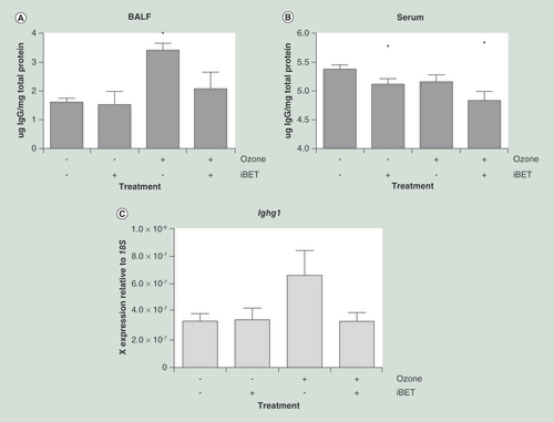
(A) Antibody titer in BALF was significantly increased by ozone exposure in acutely exposed mice (n = 8 in each group). This increase was prevented by the treatment of the mice with BET inhibitor IBET151 (30 mg/kg). (B) Antibody titer in serum was not significantly altered in serum; however, serum antibody levels were significantly reduced by IBET151 treatment. (C) Immunoglobulin gene expression (Ighg1) in whole mouse lung tissue was increased following ozone exposure, indicating that the increase in immunoglobulin titer is transcriptionally controlled. All data are shown as the mean ± the standard error.
*p < 0.05 relative to air and the placebo group.
BALF: Bronchoalveolar lavage fluid; BET: Bromo and extraterminal domain.
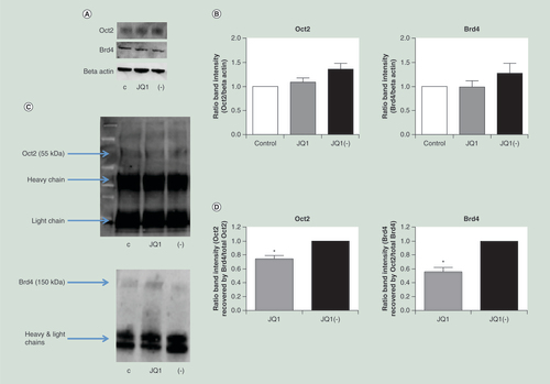
Neither Oct2 nor Brd4 levels were significantly reduced following treatment with JQ1. Proteins were measured by (A) western blot and (B) quantified (n = 3). Brd4 and Oct2 were both detectable by western blot following immunoprecipitation of either protein. (C) A representative western blot of Oct2 and Brd4 protein detected following immunoprecipitation with anti-Brd4 and anti-Oct2 antibodies respectively. (D) JQ1, compared with JQ1(-), significantly reduced the amount of Brd4 recovered following Oct2 immunoprecipitation and vice versa (n = 3). C = control, + = JQ1, - = JQ1(-). All data are shown as the mean ± the standard error.
*p < 0.05.
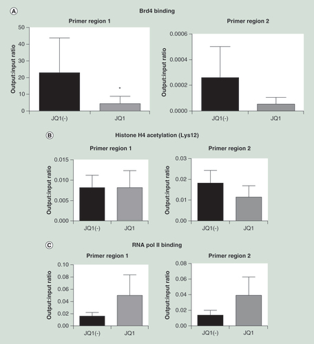
ChIP was carried out using an (A) anti-Oct2 antibody, comparing cells treated with JQ1 versus JQ1(-) controls. PCR of two putative Oct2 binding sites in the IGK promoter were carried out (n = 4). In addition, ChIP was carried out using (B) antiacetylated histone 4 and (C) RNA polymerase 2 antibodies. Data were analyzed using the Wilcoxon matched-pairs signed rank test, as data were nonparametric paired samples. All data are shown as the mean ± the standard error, *p < 0.05.
ChIP: Chromatin immunoprecipitation.