Figures & data
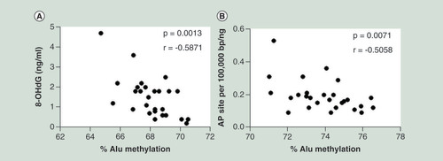
(A) The 8-hydroxy-2′-deoxyguanosine and (B) abasic sites were linearly correlated with Alu methylation levels in peripheral blood cells of healthy people. The displayed p-values are from the Pearson’s correlation test.
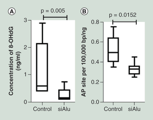
8-OHdG: 8-Hydroxy-2′-deoxyguanosine; AP site: Abasic site; r: Pearson’s correlation coefficient.
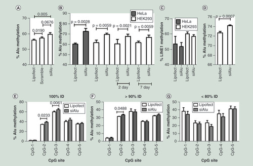
(A) The percentage of Alu methylation level in HEK293 cells transfected with only Lipofectamine reagent (lipofect), negative control siRNA from Bioneer (Scramble) and Alu siRNA (100 nM). (B) The percentage of Alu methylation level in HeLa (gray) and HEK293 (white) cells transfected with Alu siRNA. (C) The percentage of LINE-1 methylation level in HeLa (gray) and HEK293 (white) cells transfected with Alu siRNA. (D) The percentage of Alu methylation level in periodontal ligament fibroblast (passage 10) transfected with Alu siRNA. (E–G) The percentage of Alu methylation level in Alu siRNA transfected HEK293 and control (lipofect) of five CpG sites categorized by % identity to siRNA sequence (%ID) including 100% ID (E), >90% ID (F) and <80% ID (G).
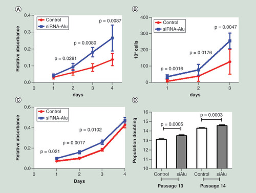
After transfection, cells were incubated for 48 h. Then, the transfected cells were seeded into 96-well plates for proliferation evaluation. (A & B) Cell proliferation in HEK293 was assessed by MTT over 4 days after Alu siRNA transfection (A) and absolute cell number was counted after seeding Alu siRNA transfected cells over 3 days (B). (C & D) Cell proliferation in periodontal ligament cells was assessed by MTT over 4 days after Alu siRNA transfection (C) and population doublings of Alu siRNA transfected cells were calculated at passage 13 and 14.
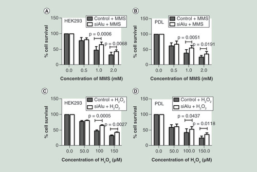
After a significant increase in Alu methylation level confirmation, control and Alu siRNA transfected cells were exposed to the increasing concentration of methyl methanesulphonate for 1 h and H2O2 for 24 h. Then growth was assessed by MTT after 48 h. Error bars indicates ± SD. (A & B) Methyl methanesulphonate sensitivity of HEK293 (A) and periodontal ligament (B). (C & D) H2O2 sensitivity of HEK293 (C) and periodontal ligament (D).
The level of 8-hydroxy-2′-deoxyguanosine (ng/ml) (A) and abasic site (per 100,000 bp/ng DNA) (B) was significantly increased in cells with Alu siRNA transfection.
8-OHdG: 8-Hydroxy-2′-deoxyguanosine; AP site: Abasic site.