11,089
Views
0
CrossRef citations to date
0
Altmetric
Review

BCOR Involvement in Cancer

, , , , &
Pages 835-855 | Received 08 Nov 2018, Accepted 01 Apr 2019, Published online: 31 May 2019

Figures & data

Figure 1. Structure and functional domains of BCOR, including BCL-6- and MLLT3-binding domains, ANK repeats and the PUFD domain.

A schematic representation of the exon structure is also shown below the protein domains.

Figure 1. Structure and functional domains of BCOR, including BCL-6- and MLLT3-binding domains, ANK repeats and the PUFD domain.A schematic representation of the exon structure is also shown below the protein domains.

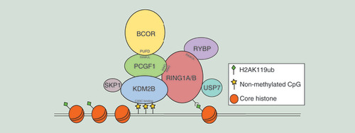
Table 1. PRC 1.1 complex components.

Figure 2. Schematic representation of the polycomb repressive complex 1.1 model.

The core complex is composed of the catalytic enzyme RING1A/B that forms a dimer with PCGF1 through the RING finger domains, and that deposits an ubiquitin moiety to histone H2A at Lys119 (H2AK119ub). BCOR binds to PCGF1 by means of its PUFD domain, while RYBP is bound to the RAWUL domain of RING1A/B. Recruitment to chromatin is due to KDM2B that recognizes nonmethylated CpG islands by its CXXC-binding domain. Other members of the complex are SKP1, that associates with KDM2B, and USP7, acting as a deubiquitinating enzyme.

PUFD: PCGF Ub-like fold discriminator.

Figure 2. Schematic representation of the polycomb repressive complex 1.1 model.The core complex is composed of the catalytic enzyme RING1A/B that forms a dimer with PCGF1 through the RING finger domains, and that deposits an ubiquitin moiety to histone H2A at Lys119 (H2AK119ub). BCOR binds to PCGF1 by means of its PUFD domain, while RYBP is bound to the RAWUL domain of RING1A/B. Recruitment to chromatin is due to KDM2B that recognizes nonmethylated CpG islands by its CXXC-binding domain. Other members of the complex are SKP1, that associates with KDM2B, and USP7, acting as a deubiquitinating enzyme.PUFD: PCGF Ub-like fold discriminator.
Figure 3. Schematic representation of different BCOR alterations, including internal tandem duplications and chimeric fusion transcripts.

The numbers inside the boxes represent the exons, while open boxes indicate UTR regions. (A) ITD–BCOR, (B) BCOR–CCNB3, (C) BCOR–MAML3, (D) ZC3H7B–BCOR and (E) BCOR–RARA.

ITD: Internal tandem duplication.

Figure 3. Schematic representation of different BCOR alterations, including internal tandem duplications and chimeric fusion transcripts.The numbers inside the boxes represent the exons, while open boxes indicate UTR regions. (A) ITD–BCOR, (B) BCOR–CCNB3, (C) BCOR–MAML3, (D) ZC3H7B–BCOR and (E) BCOR–RARA.ITD: Internal tandem duplication.

Table 2. 
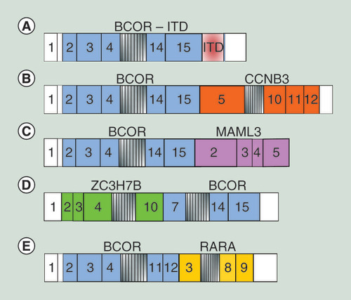
BCOR mutations in different tumor hystotypes.