Figures & data
Table 1. Most common commercially available monoclonal PD-L1 antibodies for immunohistochemical analysis to assess the expression of PD-L1 considering US FDA approvals for non-small-cell lung cancer.
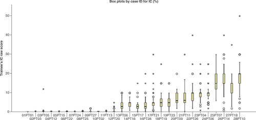
Box plot by case ID for immune cell percentage. PD-L1 (SP142) assay triple-negative breast cancer mean proficiency trainee test scores compared with expert consensus scores and case-level agreement. Scores for each case were averaged across a total of 83 pathologists who attended the training program. The median for each dataset is indicated by the black center line, and the first and third quartiles are the edges of the box area, which is known as the IQR. The extreme values (within 1.5-times the IQR from the upper or lower quartile) are the ends of the lines extending from the IQR (circles). Points at a greater distance from the median than 1.5-times the IQR are plotted individually as asterisks. These points represent potential outliers.
IC: Immune cell; IQR: Interquartile range.