Figures & data
Table 1. Molecular classification of hepatocellular adenomas.
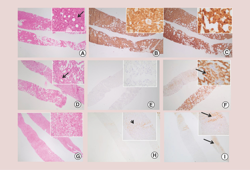
β-catenin exon-3-mutated hepatocellular adenoma: thick trabeculae with mild nuclear atypia and pseudo-gland (arrow) are frequently observed (A). Strong diffuse staining for glutamine synthase (B), as well as nuclear translocation of β-catenin (C), when present, enable diagnosis. Inflammatory hepatocellular adenoma with sinusoidal dilatations, inflammatory foci and isolated arteries (arrow) (D). No glutamine synthase expression is observed (E). Immunostaining with amyloid A (SAA) shows strong cytoplasmic staining; normal adjacent liver in the upper part is negative for SAA (F). HNF1A-inactivated adenoma with tumor steatosis (G). Glutamine synthase (H) is negative or weak (similar to adjacent nontumor liver with perivenular staining, arrow). In contrast to the adjacent nontumor liver (I, arrows), HNF1a-inactivated adenoma characteristically lacks L-FABP staining in the tumor (I).
SAA: Serum amyloid A.
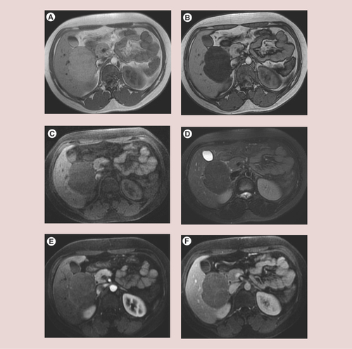
Large HNF1A-inactivated hepatocellular adenoma (HCA) in a 26-year-old woman. In chemical shift T1-weighted sequence, HCA has moderate hyperintensity in-phase (A), with a marked signal dropout on opposed-phase images (B). The lesion is hypointense in both T1 (C) and T2 (D) fat-suppressed sequences due to the lipid content. After contrast medium injection, there is only slight arterial enhancement (E), but the lesion remains hypointense compared with the normal liver on portal venous phase (F).
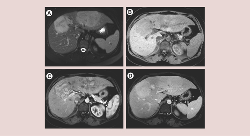
Two large inflammatory hepatocellular adenomas in a 33-year-old woman. Note: typical MRI appearance with marked hyperintensity on T2-weighted images (A), together with hypointensity on T1-weighted fat-suppressed images (B). After contrast medium injection, there is strong heterogeneous arterial enhancement of the two lesions (C) that persists in the portal venous phase (D).
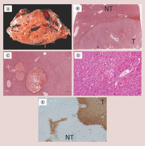
Sonic hedgehog hepatocellular adenoma: macroscopic view of a large liver nodule with hemorrhagic areas (A). Low magnification demonstrating a sharply limited hepatocellular nodule with hemorrhagic areas (B) and (C). β-catenin exon-3-mutated HCA with cellular atypia: microscopic examination shows a well-differentiated hepatocellular tumor with cellular atypia (large, hyperchromatic nuclei and bi-nucleated cells) (D). Glutamine synthase is strongly expressed in the tumor (E).
HCA: Hepatocellular adenoma; NT: Nontumor; T: Tumor.
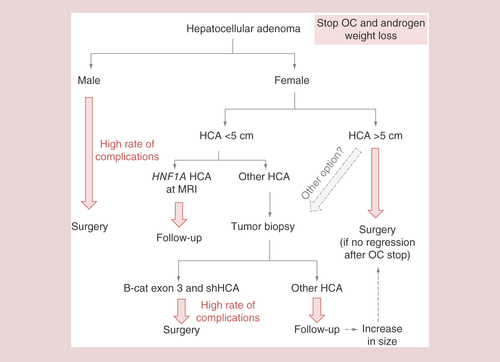
We propose an algorithm for treatment of HCA based on our knowledge of molecular classification.
HCA: Hepatocellular adenoma; OC: Oral contraception; shHCA: Sonic hedgehog hepatocellular adenoma.