Figures & data
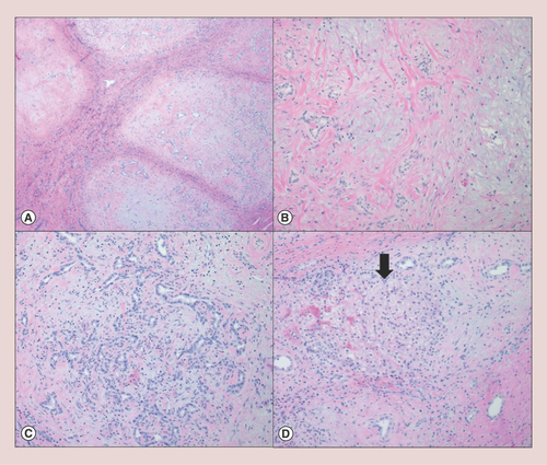
Hepatic mesenchymal hamartoma is histologically characterized by (A) a multinodular growth of myxomatous mesenchymal stroma with intervening fibrous septa (hematoxylin & eosin, 4×); (B) each nodule consists of a bland spindle cell proliferation with scattered malformed bile ducts (hematoxylin & eosin, 10×). (C) Some of the nodules demonstrate a florid bile duct proliferation with scattered lymphocytic infiltrate (hematoxylin & eosin, 10×). (D) Entrapped island of hepatocytes (arrow) is occasionally identified in the periphery of the lesion (hematoxylin & eosin, 10×).
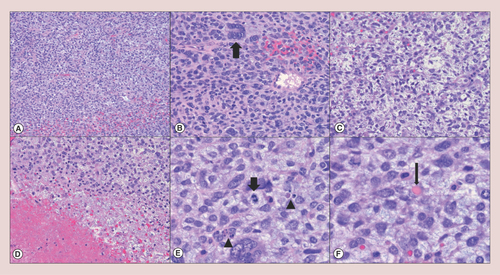
Undifferentiated embryonal sarcoma of the liver generally shows (A) hypercellular sheets of tumor cells (hematoxylin & eosin, 4×); (B) the neoplastic cells are highly pleomorphic with an increased nuclear to cytoplasmic ratio and hyperchromatic nuclei; tumor giant cells (thick arrow) are frequently identified (hematoxylin & eosin, 10×). (C) The lesional cells are focally elongated/spindled with a loose, myxoid stroma (hematoxylin & eosin, 10×). (D) Necrosis (lower left) is occasionally seen (hematoxylin & eosin, 10×). (E) Mitotic figures (thick arrow) and apoptotic bodies (arrowheads) are readily identified (hematoxylin & eosin, 20×). (F) Eosinophilic hyaline globules (thin arrow), which are PAS-positive and diastase-resistant, may be observed in the neoplastic cell cytoplasm and extracellular matrix (hematoxylin & eosin, 20×).