Figures & data
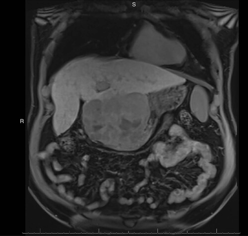
MRI: Magnetic resonance imaging.
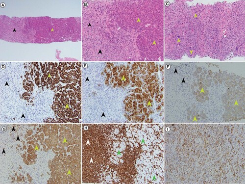
(A) At low magnification, the biphenotypic histology of sarcomatoid and conventional hepatocellular components is evident in the patient’s core needle biopsy specimen (green arrow head – conventional HCC component; black arrowhead – sarcomatoid component) (H&E 40× magnification). (B) Higher magnification shows widened plates of neoplastic hepatocytes (green arrowheads) and adjacent areas of spindled and bizarrely shaped sarcomatoid cells (black arrowheads) (H&E 100× magnification). (C) Higher magnification of sarcomatoid component with bizarre multinucleated giant cells (green arrowheads) (H&E 200× magnification). (D) The conventional HCC component (green arrowheads) is positive for HepPar1 IHC stain but the sarcomatoid component (black arrowheads) is negative (100× magnification). (E) The conventional HCC (green arrowheads) is positive for arginase 1 IHC stain but the sarcomatoid component (black arrowheads) is negative (100× magnification). (F) The conventional HCC (green arrowheads) is positive for cytokeratin CAM5.2 IHC with patchy positivity seen in the sarcomatoid component (black arrowheads) (100× magnification). (G) The conventional HCC is positive for cytokeratin AE1/AE3 IHC (green arrowheads) with patchy positivity seen in the sarcomatoid component (black arrowheads) (100× magnification). (H) The absence of staining for vimentin in the conventional HCC component (green arrowheads) stands in sharp contrast to the strong positivity of the sarcomatoid component (white arrowheads) (100× magnification). (I) Patchy positivity for cytokeratin AE1/AE3 present in this area of tumor only displaying sarcomatoid features is seen (100× magnification).
H&E: Hematoxylin and eosin; HCC: Hepatocellular carcinoma; IHC: Immunohistochemical.
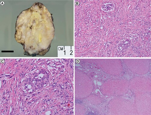
(A) A gross photograph of the resection specimen shows a variegated multinodular tumor with firm cream colored areas, yellow areas of necrosis that comprise about 25% of the tumor and areas of hemorrhage. Focal background cirrhotic liver is seen at the bottom right of the image. (B) Histologic examination of the patient’s resection specimen confirmed a predominantly sarcomatoid neoplasm with focal areas of conventional hepatocellular differentiation (H&E 100× magnification). (C) Higher magnification shows neoplastic epithelioid tumor cells surrounded by malignant spindled sarcomatoid cells (H&E 200 × magnification). (D) Cirrhotic background hepatic parenchyma (H&E × 40 magnification).