Figures & data
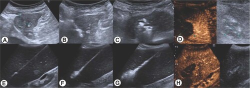
Contrast enhanced ultrasound was performed just a few minutes after the procedure and before it was finished to certify a complete ablation in the same act (A–D) HCC in close relationship with the heart. (E–H) Spleen diffuse ablation in malignant splenomegaly.
(A & B) Ultrasound CT fusion allowed a real-time synchronization of a recent imaging test where the target was clearly identified with the intraoperative ultrasound (C & D) Expert CT used for the approach of the lesion as well as for the final check of the procedure.
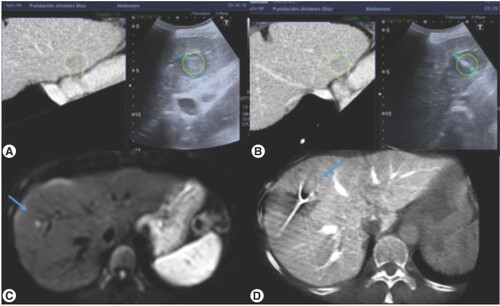
Table 1. Sample description and tumor response.
(A & B) Liver abscess after ablation of a small metastases near the hepatic hilum that was resolved with percutaneous drainage. (C & D) Tumor progression of metastases adjacent to hepatic dome vein. (E & F) Tomography performed after the introduction of lipiodol with a small amount of alcohol through the internal hole of the ablation electrode. There was a small leakage (arrow) that conditioned marked atrophy of the left lobe (star).
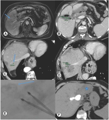
Table 2. Tumor response in primary and metastatic liver cancer.
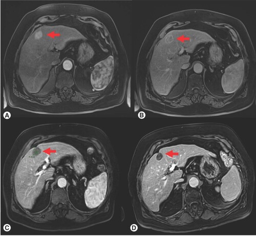
(A) Pretreatment, arterial phase on dynamic MRI, (B) Pretreatment, portal phase on dynamic MRI; (C) Post-treatment, 3-month follow-up; (D) Post-treatment, 6-month follow-up, there is a clear progressive reduction in size of the lesion as well as absence of contrast enhancement. Red arrow indicates the target liver lesion.