Figures & data
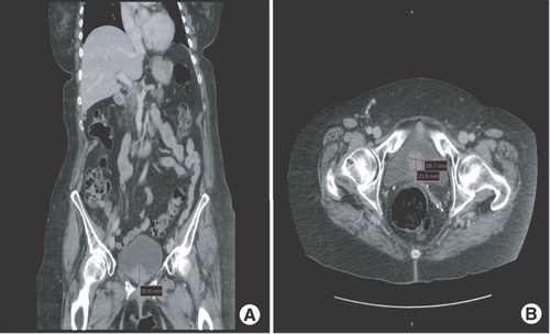
(A & B) CT intravenous pyelogram (coronal and axial views) showing polypoid soft tissue mass at the lower aspect of the urinary bladder measuring 3.3 × 3.1 × 2.4 cm compatible with the biopsy-proven malignant melanoma.
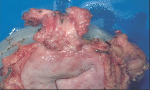
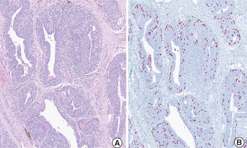
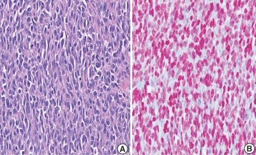
(A & B) Robotic-assisted laparoscopic radical cystourethrectomy showing sheets of highly atypical dyscohesive melanoma cells in the urinary bladder 10× (left), tumor cells demonstrate positivity for SOX10 (right).
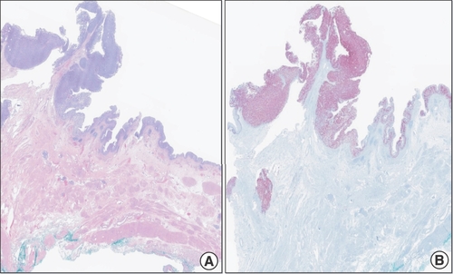
(A & B) Robotic-assisted laparoscopic radical cystourethrectomy showing the pagetoid spread of melanoma to the urethra (left), tumor cells demonstrate positivity for SOX10 (right).
(A & B) Robotic-assisted laparoscopic radical cystourethrectomy showing primary malignant melanoma of the urinary bladder with melanoma in-situ (left), specimen stains positive for SOX 10 (right).
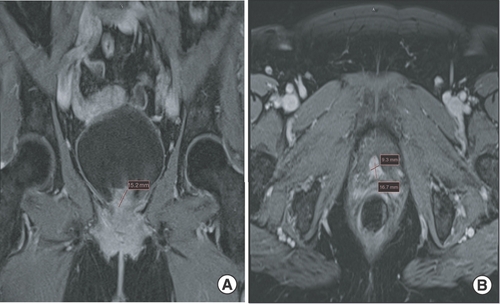
(A & B) MRI pelvis (coronal and axial views) showing nodular enhancing soft tissue mass at the posterior segment of the urethra and urinary bladder base measuring 1.7 × 1.5 × 0.9 cm compatible with the biopsy-proven malignant melanoma.
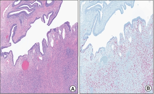
(A & B) Radical cystourethrectomy showing atypical melanoma cells of the urinary bladder with pagetoid spread of melanoma to the distal urethral margin.5× (left), tumor cells demonstrate positivity for SOX10 (right).
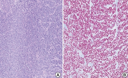
(A & B) Radical cystourethrectomy showing highly atypical epithelioid melanoma cells of the urinary bladder 40× (left), tumor cells demonstrate positivity for SOX10 (right).
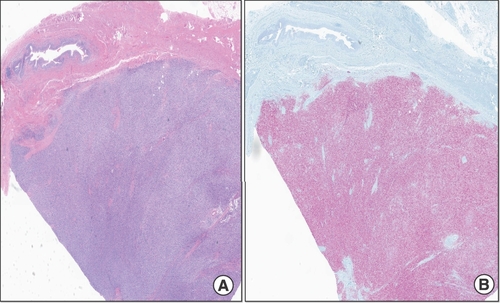
(A & B) Radical cystourethrectomy showing atypical melanoma cells in the urinary bladder diffusely involving the submucosa and muscularis propria with corresponding urothelium uninvolved 1× (left), tumor cells demonstrate positivity for SOX10 (right).