Figures & data
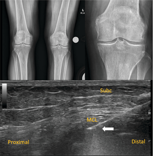
Initial evaluation where a 2.4 mm calcification is noted at the left femoral medial condyle (white arrow), also showed lateral compartment chondrocalcinosis and mild osteoarthritis (upper panel). Initial diagnostic musculoskeletal ultrasound imaging of the medial knee in a longitudinal view showing a lobulated calcium at the medial femoral condyle within the proximal medial collateral ligament. White arrow indicated the calcification of the MCL (lower panel).
Subc: Subcutaneous tissue; MCL: Medial collateral ligament; MFC: Medial femoral condyle.
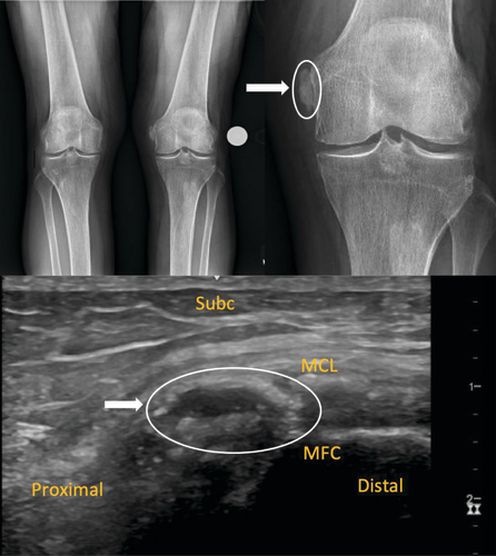
Plain radiographs AP from the 2 week-post procedure appointment showing resolution of the calcification at the medial femoral condyle with persisting lateral compartment chondrocalcinosis and mild osteoarthritis (upper panel). Ultrasound examination in long-axis to the medial collateral ligament during the TenJet™ procedure showing the needle tip (white arrow) penetrating the calcium within the MCL with decreased lucency of the calcium and reduction in size of the calcium (lower panel).
MCL: Medial collateral ligament; Subc: Subcutaneous tissue.