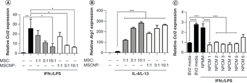
Figures & data
Mouse BV-2 microglia cells were stimulated with (A) IFN-γ and lipopolysaccharide (proinflammatory phenotype) or (B) IL-4 and IL-13 (proregenerative phenotype) either alone or with an increasing ratio of human MSCs or MSC-NPs to BV-2 cells. Relative mRNA levels of (A) mouse Ccl2 and (B) mouse Arg1 in BV-2 cells were determined by qPCR. (C)Ccl2 gene expression in BV-2 cells stimulated with IFN-γ and lipopolysaccharide either alone (BV2 media control), in the presence of unconditioned medium (NPMM), or in the presence of conditioned medium (NPCM) from six individual MSC-NP cell lines (NPCM1–6). All data are representative of at least two experiments. Values represent mean ± standard deviation.
*p < 0.05; ***p < 0.001; ****p < 0.0001.
MSC-NP: Mesenchymal stem cell-derived neural progenitor; NPCM: Neural progenitor-conditioned medium; NPMM: Neural progenitor maintenance medium.
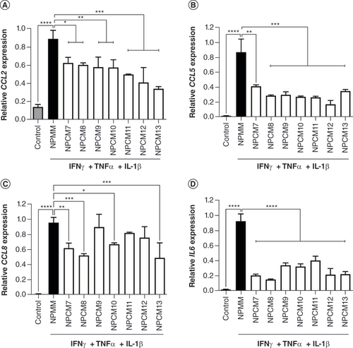
Human induced pluripotent stem cell-derived microglia cells were either unstimulated (control) or stimulated with IFN-γ, TNF-α and IL-1β (proinflammatory phenotype) in either unconditioned medium (NPMM) or MSC-NP-conditioned media (NPCM) from seven individual MSC-NP cell lines (NPCM7–13). Relative mRNA levels of proinflammatory markers (A)CCL2, (B)CCL5, (C)CCL8 and (D)IL6 were determined by qPCR. All data are representative of at least two experiments. Values represent mean ± standard deviation.
*p < 0.05; **p < 0.01; ***p < 0.001; ****p < 0.0001.
MSC-NP: Mesenchymal stem cell-derived neural progenitor; NPCM: Neural progenitor-conditioned medium; NPMM: Neural progenitor maintenance medium.
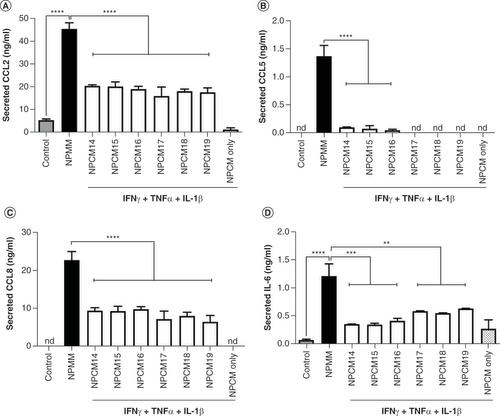
Human induced pluripotent stem cell-derived microglia cells were either unstimulated (control) or stimulated with IFN-γ, TNF-α and IL-1β in either unconditioned medium (NPMM) or MSC-NP conditioned media (NPCM) from six individual MSC-NP cell lines (NPCM14–19). Concentrations of secreted (A) CCL2, (B) CCL5, (C) CCL8 and (D) IL-6 were determined by Luminex assay. ‘NPCM only’ represents the level of secreted protein in an undiluted representative MSC-NP-conditioned medium sample. All data are representative of at least two experiments. Values represent mean ± standard deviation.
**p < 0.01; ***p < 0.001; ****p < 0.0001.
MSC-NP: Mesenchymal stem cell-derived neural progenitor; nd: Not detected; NPCM: Neural progenitor-conditioned medium; NPMM: Neural progenitor maintenance medium.
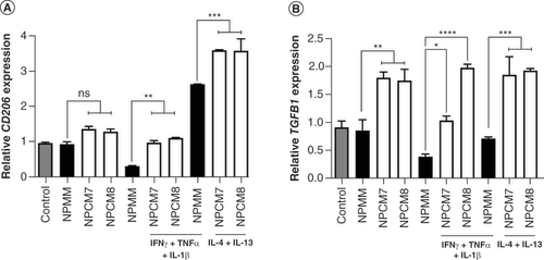
Human induced pluripotent stem cell-derived microglia cells were either unstimulated (resting phenotype), stimulated with IFN-γ, TNF-α and IL-1β (proinflammatory phenotype), or stimulated with IL-4 and IL-13 (proregenerative phenotype) in either unconditioned medium (NPMM) or MSC-NP-conditioned media from two individual MSC-NP cell lines (NPCM7 and NPCM8). Relative mRNA levels of M2 markers (A)CD206 and (B)TGFB1 were determined by qPCR. All data are representative of at least two experiments. Values represent mean ± standard deviation.
*p < 0.05; **p < 0.01; ***p < 0.001; ****p < 0.0001.
MSC-NP: Mesenchymal stem cell-derived neural progenitor; NPCM: Neural progenitor-conditioned medium; NPMM: Neural progenitor maintenance medium; ns: Not significant.
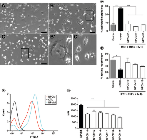
(A–C) Representative light microscopy images of microglia that were (A) unstimulated, or stimulated with IFN-γ, TNF-α and IL-1β in the presence of (B) unconditioned medium (NPMM) or (C) MSC-NP-conditioned medium. Boxed areas in (B) and (C) are shown at higher magnification in (B’) and (C’), respectively. Scale bar = 100 μm. (D & E) Quantitation of (D) activated and (E) resting morphology of stimulated microglia in the presence of MSC-NP-conditioned media from three individual MSC-NP cell lines (NPCM15, NPCM17 and NPCM19). (F) Representative histograms of microglia cultured in either NPMM or NPCM following phagocytosis of fluorescent beads. Control cells were cultured in NPMM without beads. (G) Quantitation of mean fluorescence intensity of microglia in the absence or presence of NPCM from six individual MSC-NP cell lines. Values represent mean ± standard deviation.
*p < 0.05; ***p < 0.001.
CTL: Control; MFI: Mean fluorescence intensity; MSC-NP: Mesenchymal stem cell-derived neural progenitor; NPCM: Neural progenitor-conditioned medium; NPMM: Neural progenitor maintenance medium.
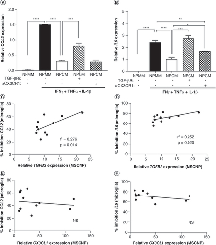
(A & B) Stimulated induced pluripotent stem cell-derived microglia either alone (NPMM), in the presence of MSC-NP-conditioned medium (NPCM25), or NPCM25 along with TGF-β receptor inhibitor (TGF-βRi), or neutralizing antibody to the CX3CL1 receptor CX3CR1 (αCX3CR1). Gene expression levels of (A)CCL2 and (B)IL6 in microglia were determined by qPCR. Values represent mean ± standard deviation. (C–F) The percentage inhibition of microglial gene expression of (C & E)CCL2 and (D & F)IL6 by NPCM samples in Figures 2 & 3 (NPCM7–19) were compared with the relative levels of (C & D)TGFB3 and (E & F)CX3CL1 gene expression in the MSC-NPs used to generate the conditioned media. MSC-NPs for NPCM14 and NPCM18 were omitted due to lack of available RNA.
*p < 0.05; **p < 0.01; ***p < 0.001; ****p < 0.0001.
MSC-NP: Mesenchymal stem cell-derived neural progenitor; NPCM: Neural progenitor-conditioned medium; NPMM: Neural progenitor maintenance medium; NS: Not significant.