Figures & data
Figure 1. Case 1. A 60-year-old woman presented with slowly progressive bilateral hearing loss from age 40. By age 50 she had only minimal gain from hearing aids and when we first saw her they were nearly useless in her daily life. COMBI40+ with regular electrode was used for this patient on Dec 10, 2008. For insertion, the round window approach was applied, and full insertion was achieved. Complete preservation of residual hearing was obtained. (A) Endoscopic view of round window insertion, (B) postoperative X-ray finding, (C) imaging with putative location of electrode and the referential tonotopic map, (D) preoperative and postoperative audiograms. The image of human cochlea neural tissues stained by osmium tetroxide used in was kindly provided by Dr C.G. Wright, USWT, Dallas, USA (red, mm from round window; black, corresponding frequency).
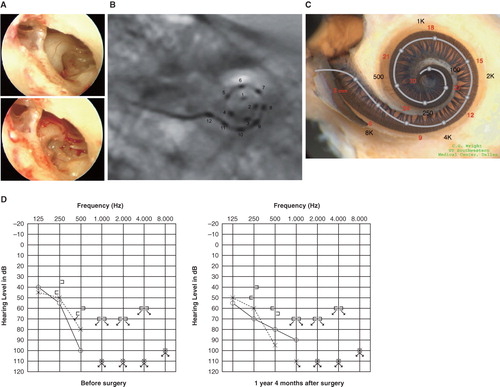
Figure 2. Case 2. This 39-year-old man was congenitally deaf in the left ear. Mild hearing loss in his right ear was noticed in childhood, and he presented with progressive hearing loss of 10 years duration. FLEXeas/RW approach was applied on Nov 16, 2009. Preservation of residual hearing was obtained. (A) Endoscopic view of round window insertion, (B) postoperative X-ray finding, (C) imaging with putative location of electrode and the referential tonotopic map, (D) preoperative and postoperative audiograms.
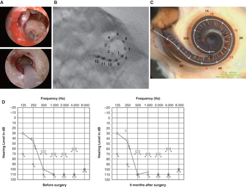
Figure 3. Case 3. This 45-year-old woman became aware of bilateral hearing loss and tinnitus around age 25. When she presented to us it had been slowly progressing for 10 years. PULSAR FLEXeas/RW approach was applied on Nov 18, 2009. Preservation of residual hearing was obtained. (A) Endoscopic view of round window insertion, (B) postoperative X-ray finding, (C) imaging with putative location of electrode and the referential tonotopic map, (D) preoperative and postoperative audiograms.
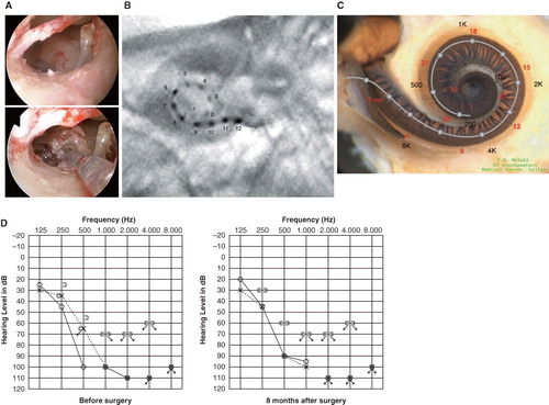
Figure 4. Case 4. This 38-year-old woman had hearing loss detected by mass screening in primary school. It appeared to slowly progress as she grew up, and by age 25 she suffered inconvenience in hearing and communication, mainly using only her left ear. The PULSAR FLEXeas/RW approach was applied on Dec 21, 2009. Preservation of residual hearing was obtained. (A) Endoscopic view of round window insertion, (B) postoperative X-ray finding, (C) imaging with putative location of electrode and the referential tonotopic map, (D) preoperative and postoperative audiograms.
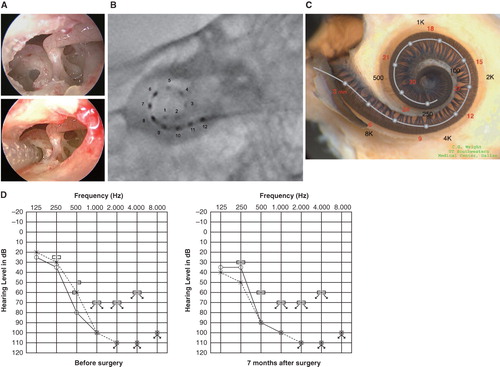
Figure 5. Case 5. This 68-year-old man presented with slowly progressive bilateral hearing loss from around age 40. He had only minimal gain from hearing aids. The PULSAR FLEXsoft/RW approach was applied on May 17, 2010. Preservation of residual hearing was obtained. (A) Endoscopic view of round window insertion, (B) postoperative X-ray finding, (C) imaging with putative location of electrode and the referential tonotopic map, (D) preoperative and postoperative audiograms.
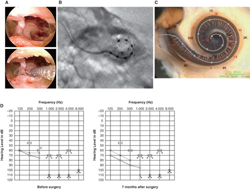
Figure 6. Video recording showing that drilling time to reach the perilymphatic space is significantly shorter for the round window approach compared with cochleostomy insertion.
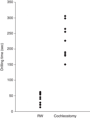
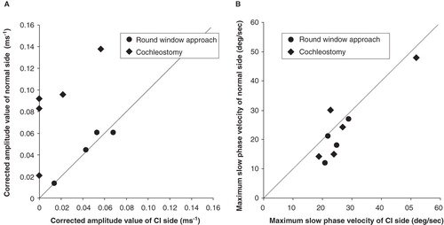
Figure 7. (A) Vestibular evoked myogenic potential (VEMP) responses were recorded in four of five cases and were well preserved postoperatively. VEMP responses decreased postoperatively in the cochleostomy cases while they were maintained in the round window insertion cases. Corrected amplitude value Cp13-n23 (ms–1) = amplitude Cp13-n23 (micro V)/background electromyographic activities (micro V ms). (B) Caloric response was well preserved and there were no differences between the two groups. MSV, maximum slow eye velocity.