Figures & data
Figure 1. Pure-tone audiograms: (A) a 22-year-old female with a GJB2 mutation; (B) a 26-year-old male with an SLC26A4 mutation. There were no clear differences in hearing thresholds in these two cases.
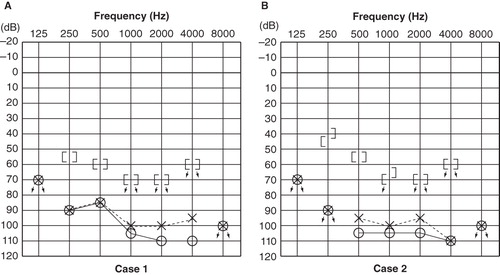
Figure 2. Transaxial PET images of each participant's brain: activation (arrowheads) of the superior temporal gyrus with visual language stimuli in each case. (A) Case 1 (GJB2 mutation). The superior temporal gyri were strongly activated bilaterally. (B) Case 2 (SLC26A4 mutation). The superior temporal gyri exhibited less activation than in case 1.
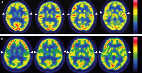
Figure 3. Cortical activation by language-related visual stimuli in the two profoundly deafened cases. Case 1 (GJB2 mutation) showed significant activation in the right middle temporal gyrus [BA21] (1), superior temporal gyrus [BA22] (2), and left superior temporal gyrus [BA42] (3), and left cerebellum (4), while case 2 (SLC26A4 mutation) exhibited significant activation in the right superior frontal gyrus [BA9] (1), and middle temporal gyrus [BA20] (2) (SPM2, p < 0.001, uncorrected).
![Figure 3. Cortical activation by language-related visual stimuli in the two profoundly deafened cases. Case 1 (GJB2 mutation) showed significant activation in the right middle temporal gyrus [BA21] (1), superior temporal gyrus [BA22] (2), and left superior temporal gyrus [BA42] (3), and left cerebellum (4), while case 2 (SLC26A4 mutation) exhibited significant activation in the right superior frontal gyrus [BA9] (1), and middle temporal gyrus [BA20] (2) (SPM2, p < 0.001, uncorrected).](/cms/asset/5a2f37eb-e197-48fd-b223-eec0db4d94ac/ioto_a_593719_f0003_b.jpg)