Figures & data
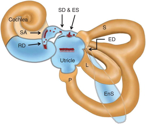
Figure 1. 3D CT image of the reuniting duct (RD), saccular duct (SD) and endolymphatic sinus (ES) of a healthy volunteer's ear (left), cadaver's ear (upper right), and schematic view (lower right). All these views are left ears. Red arrows show the RD and yellow arrows show the SD and ES. CR, cochlear recess; EnS, endolymphatic sac; IAC, internal auditory canal; L, lateral semicircular canal; OS, osseous spiral lamina; P, posterior semicircular canal; S, superior semicircular canal; SA saccule; SR, saccular recess; UR, utricular recess.
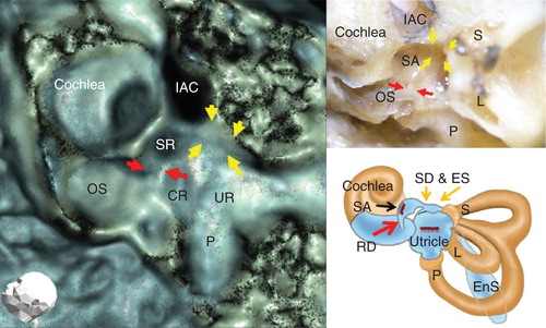
Figure 2. Drawings demonstrating the patency of the reuniting duct (RD), saccular duct (SD) and endolymphatic sinus (ES) on 3D CT images. When otoconia are dislodged into the RD, SD or ES, they lose continuity of the surface of their bony grooves and become vague on 3D CT images.
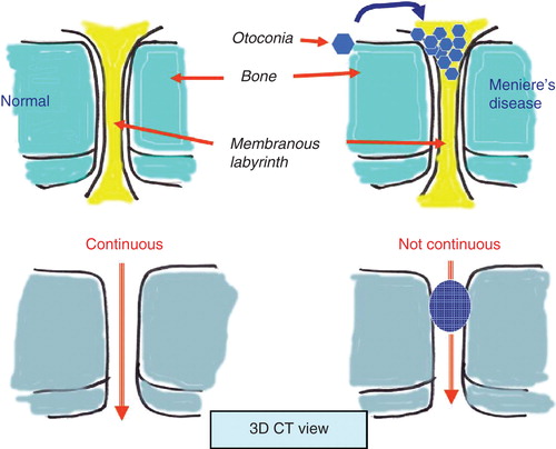
Figure 3. Representative views of the left ear of a patient with Meniere's disease (MD) (A) and a volunteer's healthy ear (B). (A) Although the outline of the bony groove of the reuniting duct (RD) (arrows), saccular duct (SD) and endolymphatic sinus (ES) can be traced, their luminal spaces seem to be occupied by a dense, bony substance. (B) On the other hand, those of the volunteer's healthy ear do not seem to be occupied by such a substance, and the luminal spaces maintain continuity. The arrowhead shows the discontinuity of the groove of the RD (arrows) in the MD patient. IAC, internal auditory canal; SR, saccular recess.
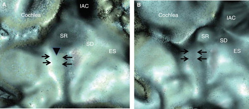
Figure 4. Representative views of the right ear of a patient with Meniere's disease (MD) (A) and a volunteer's healthy ear (B). (A) Both the bony grooves of the reuniting duct (RD) and the endolymphatic sinus (ES) are fully occupied by a dense, bony substance and it is hard to trace their luminal spaces, but that of the saccular duct (SD) is not fully occupied in the MD patient. (B) The bony grooves of the RD, SD and ES of the volunteer's healthy ear are not occupied and maintain their luminal spaces. The bony grooves of the SD and ES show continuous images (arrows). IAC, internal auditory canal; SR saccular recess.
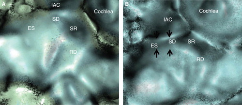
Table I. Status of the reuniting duct (RD), saccular duct (SD) and endolymphatic sinus (ES) in 62 patients with Meniere's disease (MD) and 13 healthy volunteers.
Figure 5. Distribution pattern of the reuniting duct (RD) in affected and non-affected ears of patients with Meniere's disease (MD) and healthy ears of volunteers.
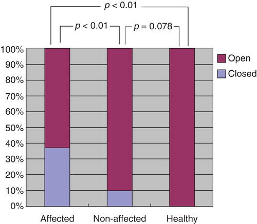
Figure 6. Distribution pattern of the saccular duct (SD) in affected and non-affected ears of patients with Meniere's disease (MD) and healthy ears of volunteers.
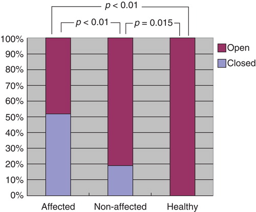
Figure 7. Distribution pattern of the endolymphatic sinus (ES) in affected and non-affected ears of patients with Meniere's disease (MD) and healthy ears of volunteers.
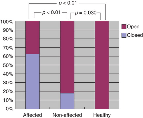
Figure 8. 3D CT views of a cadaver temporal bone. The bony grooves underlining the saccular duct (SD) and the endolymphatic sinus (ES) treated with CaCO3 (A) appear vague compared with those treated with muscle fragments (B). Note the difference in continuity of the bony grooves (arrowheads). IAC, internal auditory canal; RD, reuniting duct; SR, saccular recess.
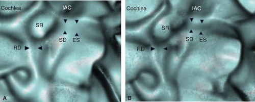
Figure 9. Hypothetical view of blockage of the reuniting duct (RD), saccular duct (SD) and endolymphatic sinus (ES) by dislodged otoconia from the saccule in patients with Meniere's disease (MD). ED, endolymphatic duct; EnS, endolymphatic sac; L, lateral semicircular canal; P, posterior semicircular canal; S, superior semicircular canal; SA, saccule.