Figures & data
Figure 1. Fundus photographs in February 2014 (A) and fluorescein angiography in April 2014 (B).
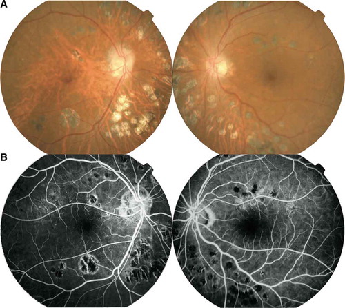
Figure 2. Optical coherence tomography images. (A) Stage 2 macular hole (arrow) in the right eye December 2007. (B) Stage 2 macular hole (arrow) in the left eye January 2013. (C) Bilateral macular holes were successfully closed after a combined vitrectomy/phacoemulsification, images taken in February 2013.
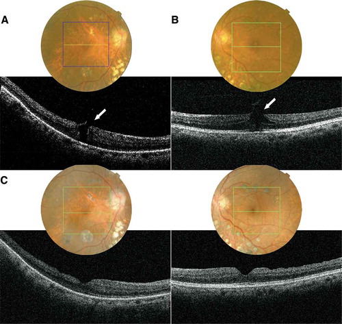
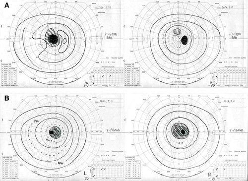
Figure 3. Goldmann visual field testings. (A) Central scotoma OS and an asymptomatic paracentral relative scotoma OD in February 2014. (B) Central scotomas OU in July 2014.