Figures & data
Figure 1. (A) Measurement set-up with 4-electrode measurement probe and LCR measurement bridge; (B) 4-electrode measurement probe in detail.
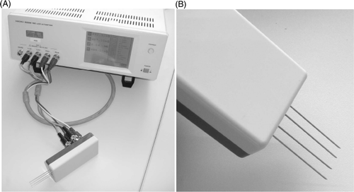
Figure 2. Experimental set-up and measurement order of ex vivo examination in porcine liver.
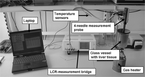
Figure 3. Experimental set-up of in vivo examinations in perfused porcine liver.
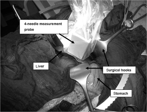
Figure 4. Measurement curve of medians from 25 individual measurements from ex vivo porcine liver (grey points, changes during the heating phase; white points, changes during the cooling phase).
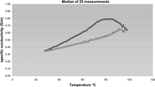
Table I. Specific conductivity during the coagulation process (median with min/max is given in steps of 5°C).
Table II. Specific conductivity during the cooling process (median with min/max is given in steps of 5°C).