Figures & data
Figure 1. Optimal thermal response of a single pulse at a new focal spot location (and at the corresponding critical normal tissue location) when the normal tissue temperature is constrained. In this example the temperature in the focal spot being heated starts at body temperature, while the normal tissue temperature starts at an elevated level due to thermal build-up accumulated by the previous pulses. The normal tissue is allowed to cool to its optimal value between pulses.
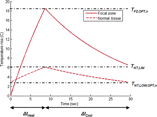
Figure 2. HIFU treatment optimisation algorithm used for calculating the heating and interpulse cooling times at a single focal spot location.
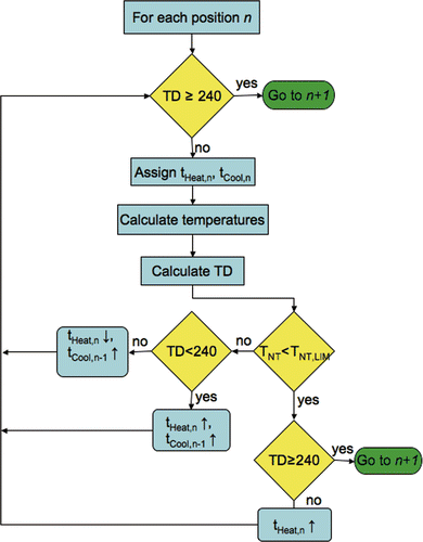
Figure 3. Schematic of 3D simulation geometries. The distances for the 1 cm3 superficial tumour geometry are: D1 = 11.2 cm, D2 = 3.3 cm, D3 = 0.3 cm. The distances for the 4.4 cm3 tumour are D1 = 7.8 cm, D2 = 10 cm, D3 = 4.6 cm. The origin is located on the longitudinal axis of the transducer. Normal tissue constraint planes are shown (dashed lines), located 1 cm above and below the treated tumour volume in both cases.
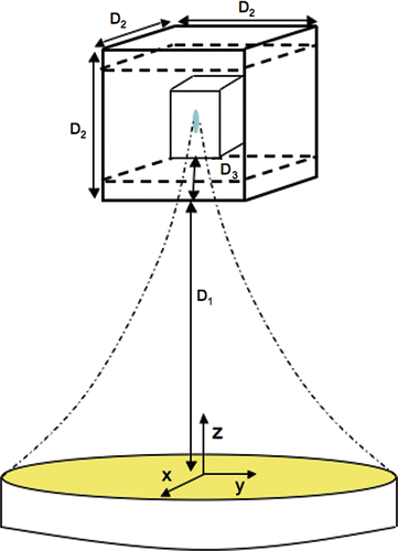
Figure 4. Simulated power density (W/m3) contours for the 1-cm3 tumour volume. Shown are the contours in a y-z plane through the transducer's longitudinal axis when the transducer's geometric focus is located in the middle treatment plane. FWHM dimensions are 2 mm × 10 mm.
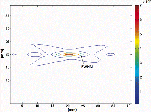
Table I. Thermal and acoustic parameters used in the simulations.
Figure 5. Scanning sequences used in the 1-cm3 superficial tumour simulations. Each sequence heated 45 focal spots in a 3 × 5 × 3 pattern: (A) x-y raster pattern, (B) y-z raster pattern, (C) alternating pattern with reduced normal tissue SAR overlap. The 4.4-cm3 tumour used similar patterns with 243 points.
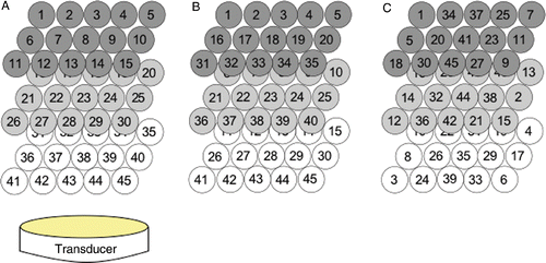
Figure 6. (A) Optimised total treatment times (ttreat) for both tumour sizes as a function of the applied power magnitude for a tissue perfusion value of 5 kg/m3s, an alternating scanning path, and a normal tissue constraint limit of 6°C above basal temperature. (B) Corresponding cumulative heating time (solid lines) and cooling time (dashed lines) for the two tumour sizes.
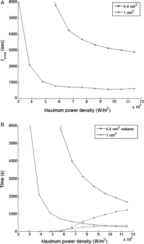
Figure 7. Optimised treatment time results for normal tissue temperature constraint limits of 6°C, 7°C and 8°C above the basal value versus total applied power for the 1-cm3 superficial tumour, a perfusion value of 0.5 kg/m3s, and the alternating scanning path trajectory.
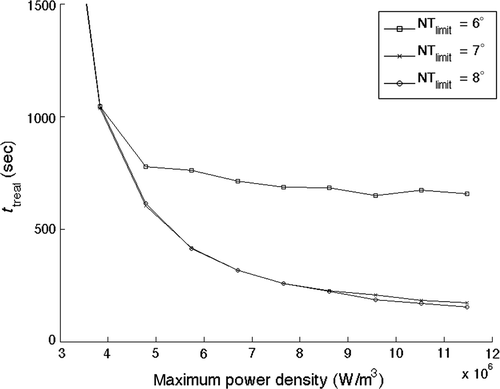
Figure 8. Comparison of optimised total treatment time for perfusion rates of 0.5, 5 and 10 kg/m3s for the alternating path in the 1-cm3 tumour geometry: (A) total treatment time, (B) cumulative heating time (solid lines) and cooling time (dashed lines).
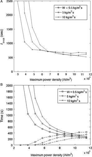
Figure 9. (A) Optimised total treatment time and (B) cumulative heating time (solid lines) and cooling time (dashed lines) versus focal zone power density for optimised HIFU treatments with a perfusion value of 5 kg/m3s for the three scanning paths in applied to the 1-cm3 tumour geometry.
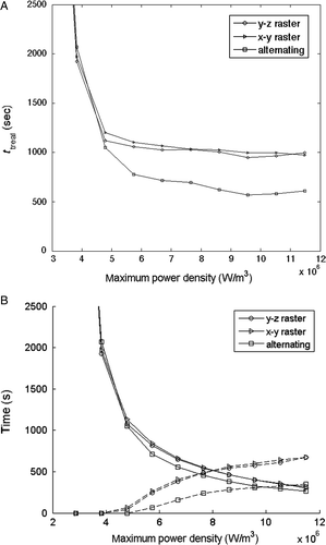
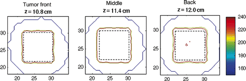
Figure 10. Thermal dose accumulated in the 4.4-cm3 tumour volume. Shown are the front (z = 10.8 cm), middle (z = 11.4 cm) and back (z = 12 cm) planes of the tumour. The tumour boundary is shown as a dashed line.