Figures & data
Figure 1. Systolic blood flow velocities measured by phase-contrast MRI in the large abdominopelvic arteries before and 30 min after RHT of the cervix. Note that flow velocity in the large pelvic arteries (iliac and femoral arteries), which give off smaller branches to the pelvic muscles, increases by 100% after hyperthermia. There is only little change in flow velocity in the inferior mesenteric artery, the superior rectal artery, and the two sigmoid arteries, which supply highly perfused organs (large intestine and rectum).
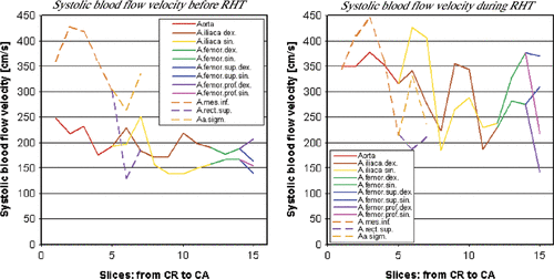
Figure 2. HT treatment in a patient with sarcoma. Morphologic image (A) and temperature map with superimposed 42°C isotherms. Also shown is the catheter for insertion of a temperature sensor (B).
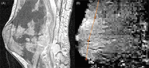
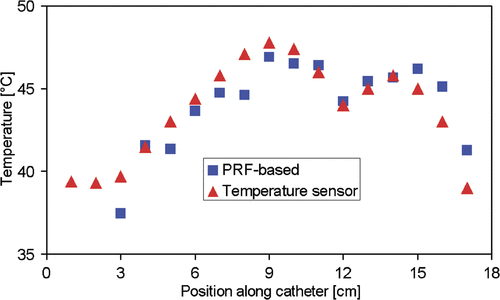
Figure 3. Comparison of the temperatures obtained using the PRFS technique with the temperatures measured by a sensor along the catheter tract in a patient with soft tissue sarcoma of the calf.