Figures & data
Figure 1. Experimental set-up featuring fluid-filled phantom with heating bath (white arrow) and thermometer with optical temperature probe (black arrow).
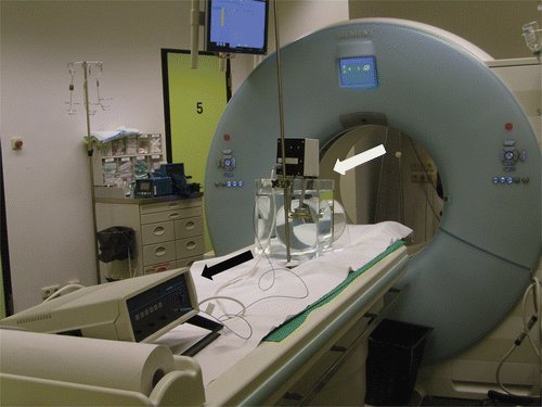
Table I. Scan parameters used for the two sets of experiments.
Table II. Least square means of the standard deviation of area averaged CT numbers depending on tube current time product at 120 kV.
Table III. Least square means of the standard deviation of area averaged CT numbers depending on tube voltage at 250 mAs.
Table IV. Least square means of the standard deviation of area averaged CT numbers depending on collimation and reconstructed slice thickness.
Table V. Results of the regression analysis (temperature versus CT number) for the different fluids.
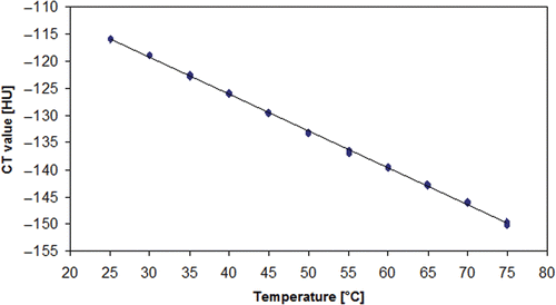
Figure 2. Regression line for the relationship between temperature of water samples and measured CT number (imaging parameters: 250 mAs, 140 kV, 1.2 mm collimation, 9.6 mm slice thickness).
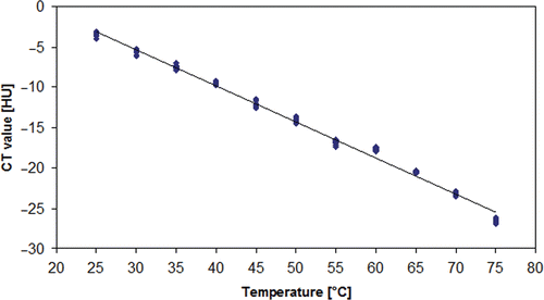
Figure 3. Regression line for the relationship between temperature of sunflower oil samples and measured CT number (imaging parameters: 250 mAs, 140 kV, 1.2 mm collimation, 9.6 mm slice thickness).