Figures & data
Figure 1. Experimental set-up for the ex vivo RFA of turkey breast muscle (not to scale). The RFA needle probe is shown.
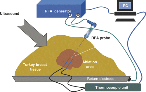
Figure 2. Experimental set-up for ex vivo CITT ablation in a turkey breast muscle (not to scale).
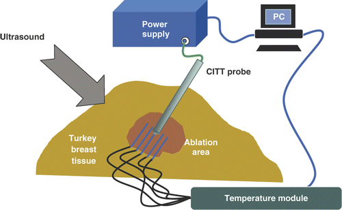
Figure 3. Example of observed colour Doppler ultrasound signals (DUS) superimposed on B-mode images. The two images shown were taken at different times during an RFA ablation experiment using the needle probe. The letter A indicates the location of the RFA probe while letters B and C denote thermocouple locations (2 mm apart as measured with the scanner).

Figure 4. Example of Doppler imaging artefacts during RFA in the form of monochromatic strips.
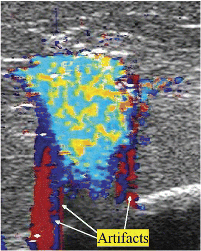
Figure 5. Thermocouple recorded temperature histories during an ex vivo RFA experiment in turkey breast muscle. The colour-coded shaded areas denote the times and temperatures of observed sustained Doppler activity for the respective thermocouples (e.g. blue shaded area corresponds to temperatures as function of time measured at the probe). The positions of the RFA probe (A) and thermocouples B and C are as shown in .
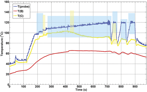
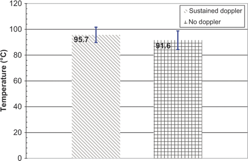
Figure 6. T1 and T2 for the eight RFA experiments. Error bars correspond to standard deviations.
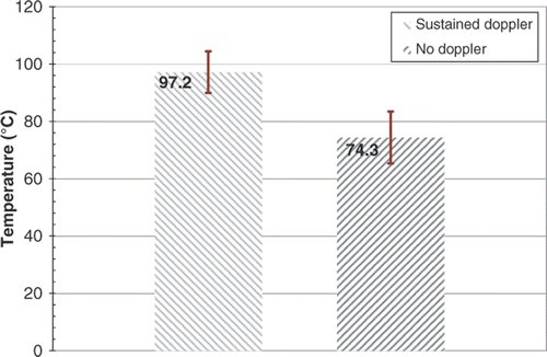
Figure 7. Example of sustained DUS at two different instances during one of the eight CITT ablation experiments. Letters B, C, D, and E denote the locations of the thermocouples with their corresponding temperatures at that time. The circular echo (pointed with red arrow) shows the position of the surface of the CITT probe at point A (10 mm in diameter).
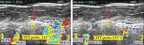
Figure 8. Thermocouple recorded temperature histories during a CITT ablation experiment in turkey breast muscle. The colour-coded shaded areas denote the times and temperatures of observed sustained Doppler activity for the respective thermocouples (e.g. blue shaded area corresponds to temperatures as function of time measured at the probe, the pink shaded area corresponds to the red line, T(B)). The locations of the CITT probe and the thermocouples B through E are as shown in .
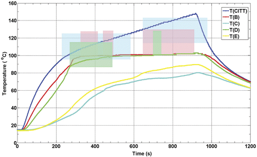
Figure 9. T1 and T2 for the eight CITT ablation experiments. Error bars correspond to standard deviations.