597
Views
27
CrossRef citations to date
0
Altmetric
Research Article

The potential role of dynamic MRI in assessing the effectiveness of high-intensity focused ultrasound ablation of breast cancer

, , MD, , , &
Pages 594-603 | Received 08 Sep 2009, Accepted 25 Mar 2010, Published online: 13 Aug 2010

Figures & data

Figure 1. Diagrams of technical effectiveness. (A) Illustration of HIFU therapy. (B) Illustration of the technical effectiveness as divided into complete versus partial ablation. (C) Illustration of the technical effectiveness as depicted on a MR subtraction image: thin rim enhancement for complete ablation and nodular or irregular thick enhancement for partial ablation.
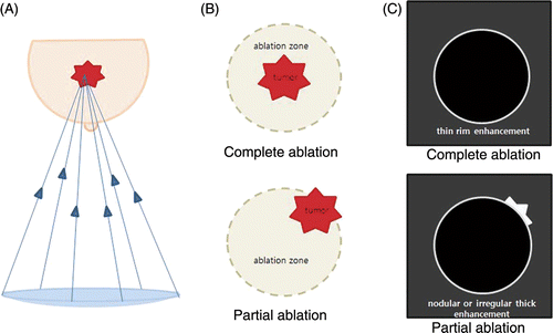
Figure 1. Diagrams of technical effectiveness. (A) Illustration of HIFU therapy. (B) Illustration of the technical effectiveness as divided into complete versus partial ablation. (C) Illustration of the technical effectiveness as depicted on a MR subtraction image: thin rim enhancement for complete ablation and nodular or irregular thick enhancement for partial ablation.

Table I.  Ablation parameters of HIFU.

Table II.  Summary of MRI findings and pathology after HIFU procedure.

Figure 2. Dynamic MRI findings of patient 1 mentioned in . (A) An early post-contrast sagittal T1-weighted image prior to HIFU showed a 12 mm, irregular enhancing mass (arrow). (B) A pre-contrast axial T1-weighted image of MRI post-HIFU revealed an isointense index tumour (arrow). (C) An early post-contrast sagittal T1-weighted image of MRI post-HIFU revealed an index tumour, isointense to adjacent parenchyma (arrow). It was surrounded by an ablation zone of decreased signal intensity (arrowheads). (D) The ablation zone on a delayed subtraction image of MRI post-HIFU was demarcated by dark signal intensity. Although no enhancement of the index tumour was observed, an incomplete thin rim enhancement was present (arrowheads). (E) An early post-contrast sagittal T1 weighted image of 12 months FU MRI, the size of the index tumour (arrow) had decreased to 5 mm. (F) The size of the ablation zone surrounded by a thin rim enhancement (arrowheads) had also decreased on a delayed subtraction image of 30 months FU MRI.
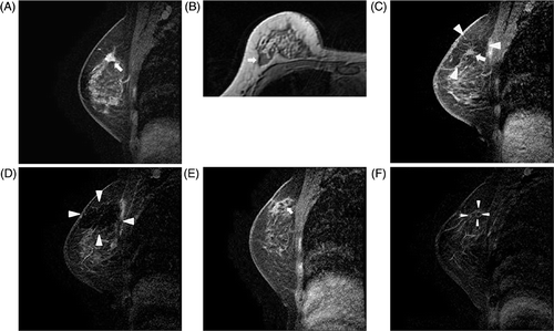
Figure 2. Dynamic MRI findings of patient 1 mentioned in Table II. (A) An early post-contrast sagittal T1-weighted image prior to HIFU showed a 12 mm, irregular enhancing mass (arrow). (B) A pre-contrast axial T1-weighted image of MRI post-HIFU revealed an isointense index tumour (arrow). (C) An early post-contrast sagittal T1-weighted image of MRI post-HIFU revealed an index tumour, isointense to adjacent parenchyma (arrow). It was surrounded by an ablation zone of decreased signal intensity (arrowheads). (D) The ablation zone on a delayed subtraction image of MRI post-HIFU was demarcated by dark signal intensity. Although no enhancement of the index tumour was observed, an incomplete thin rim enhancement was present (arrowheads). (E) An early post-contrast sagittal T1 weighted image of 12 months FU MRI, the size of the index tumour (arrow) had decreased to 5 mm. (F) The size of the ablation zone surrounded by a thin rim enhancement (arrowheads) had also decreased on a delayed subtraction image of 30 months FU MRI.

Figure 3. Partial ablation in a 68-year-old woman (patient 4 mentioned in ). (A) An early post-contrast sagittal T1-weighted image prior to HIFU showed a 15 mm irregular enhancing mass (arrow). (B) An axial fat suppressed T2-weighted image prior to HIFU revealed an isointense index tumour (arrow). (C) An axial fat suppressed T2-weighted image of MRI post-HIFU showed an isointense index tumour (arrow) with mammary oedema. (D) An early post-contrast sagittal T1-weighted image of MRI post-HIFU revealed an index tumour, isointense to adjacent parenchyma (arrow) surrounded by an ablation zone of decreased signal intensity (arrowheads). Irregular enhancement was seen at the periphery of the ablation zone (black arrow). (E) A central dark signal intensity and a nodular or irregular thick enhancement (arrow) was observed on the early and delayed subtraction image of MRI post HIFU. (F) Kinetic curve pattern showed early rapid enhancement and delayed persistent pattern. (G) Photomicrograph of a mastectomy specimen showing prevalent coagulative necrosis and a small portion of viable tumour (arrows) (H and E, original magnification × 40).
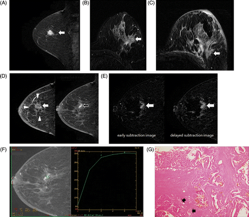
Figure 3. Partial ablation in a 68-year-old woman (patient 4 mentioned in Table II). (A) An early post-contrast sagittal T1-weighted image prior to HIFU showed a 15 mm irregular enhancing mass (arrow). (B) An axial fat suppressed T2-weighted image prior to HIFU revealed an isointense index tumour (arrow). (C) An axial fat suppressed T2-weighted image of MRI post-HIFU showed an isointense index tumour (arrow) with mammary oedema. (D) An early post-contrast sagittal T1-weighted image of MRI post-HIFU revealed an index tumour, isointense to adjacent parenchyma (arrow) surrounded by an ablation zone of decreased signal intensity (arrowheads). Irregular enhancement was seen at the periphery of the ablation zone (black arrow). (E) A central dark signal intensity and a nodular or irregular thick enhancement (arrow) was observed on the early and delayed subtraction image of MRI post HIFU. (F) Kinetic curve pattern showed early rapid enhancement and delayed persistent pattern. (G) Photomicrograph of a mastectomy specimen showing prevalent coagulative necrosis and a small portion of viable tumour (arrows) (H and E, original magnification × 40).

Figure 4. Complete ablation and mammary oedema in a 52-year-old woman (patient 3 mentioned in ). (A) On an axial fat suppressed T2-weighted image of MRI post-HIFU, mammary oedema (arrow) was accompanied by skin and trabecular thickening and thermal injury was observed as irregular hyperintensity within the chest wall muscle (arrowhead). (B) On an axial fat suppressed T2-weighted image of 6 months FU MRI, mammary oedema and chest wall muscle change almost disappeared. (C) The subtraction image of 11 months FU MRI showed a central dark signal intensity and a thin rim enhancement (arrow). Thin rim enhancement was faint on early subtraction image, but was more prominent on the delayed subtraction image. (D) Photomicrograph of mastectomy specimen shows coagulative necrosis (arrows) and peripheral foreign body reaction (arrowheads) (H and E, original magnification × 1). No viable tumour was present in the ablation zone

Figure 4. Complete ablation and mammary oedema in a 52-year-old woman (patient 3 mentioned in Table II). (A) On an axial fat suppressed T2-weighted image of MRI post-HIFU, mammary oedema (arrow) was accompanied by skin and trabecular thickening and thermal injury was observed as irregular hyperintensity within the chest wall muscle (arrowhead). (B) On an axial fat suppressed T2-weighted image of 6 months FU MRI, mammary oedema and chest wall muscle change almost disappeared. (C) The subtraction image of 11 months FU MRI showed a central dark signal intensity and a thin rim enhancement (arrow). Thin rim enhancement was faint on early subtraction image, but was more prominent on the delayed subtraction image. (D) Photomicrograph of mastectomy specimen shows coagulative necrosis (arrows) and peripheral foreign body reaction (arrowheads) (H and E, original magnification × 1). No viable tumour was present in the ablation zone

Figure 5. Partial ablation in a 60-year-old woman (patient 5 mentioned in ). (A) A central dark signal intensity and a nodular or irregular thick enhancement (arrow) was observed on the early subtraction image of MRI post HIFU. (B) Kinetic curve pattern showed early rapid enhancement and delayed washout pattern.
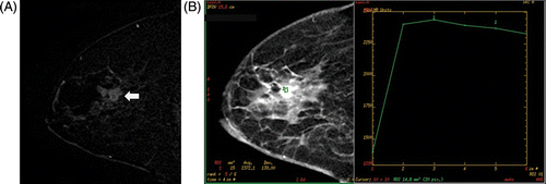
Figure 5. Partial ablation in a 60-year-old woman (patient 5 mentioned in Table II). (A) A central dark signal intensity and a nodular or irregular thick enhancement (arrow) was observed on the early subtraction image of MRI post HIFU. (B) Kinetic curve pattern showed early rapid enhancement and delayed washout pattern.

Reprints and Corporate Permissions

Please note: Selecting permissions does not provide access to the full text of the article, please see our help page How do I view content?

To request a reprint or corporate permissions for this article, please click on the relevant link below:

Academic Permissions

Please note: Selecting permissions does not provide access to the full text of the article, please see our help page How do I view content?

Obtain permissions instantly via Rightslink by clicking on the button below:

If you are unable to obtain permissions via Rightslink, please complete and submit this Permissions form. For more information, please visit our Permissions help page.