Figures & data
Figure 1. Changes in the (A) glucose (Glc) concentration, (B) weight and (C) fluorescence intensity (F%) of advanced glycated end products (AGEs) during the time course of the experiment in different groups of rats. In all figures (except ), the same symbols are used. ♦ Group 1 Control Room Temperature Air, ▪ Group 2 Diabetic Room Temperature Air, ▴ Group 3 Control group with HTT and □ Diabetic group with HTT. Data are means ± SD (n = 5–7). *P < 0.05, **P < 0.01 and ***P < 0.001 show the significant changes between two diabetic groups with or without HTT. ♣ shows the borderline of significance (0.05 < p < 0.1) in weight and glucose (P = 0.077 and P = 0.062, respectively).
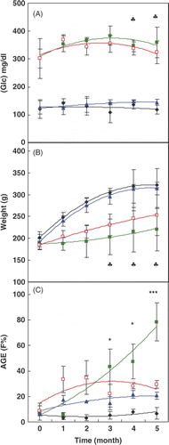
Table I. Comparison of serum insulin levels in control and diabetes rats with or without hot-tub therapy (HTT).
Figure 2. Changes in the lipid profile, including: (A) triglyceride (TG), (B) total cholesterol (TC), (C) high density lipoprotein cholesterol (HDL-c) and (D) low density lipoprotein cholesterol (LDL-c) during the time course of the experiment in different groups of rats (the same indicators are used as in ). Data are means ± SD (n = 5–7). *P < 0.05, **P < 0.01 and ***P < 0.001 show the significant changes between two diabetic groups with or without HTT.
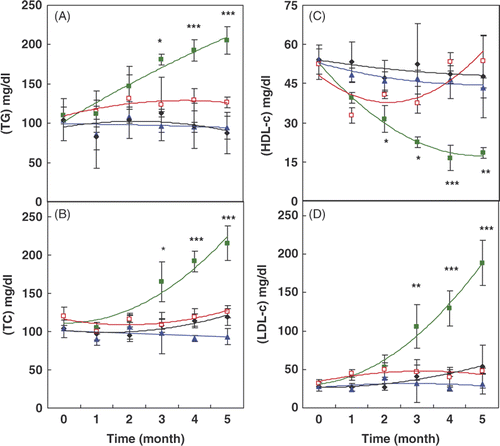
Figure 3. Changes of the ferric reducing ability of plasma (FRAP) during the time course of the experiment in different groups of rats. In this figure the same indicators are used as in . Data are means ± SD (n = 5–7). *P < 0.05, **P < 0.01 and ***P < 0.001 show the significant changes between two diabetic groups with or without HTT.
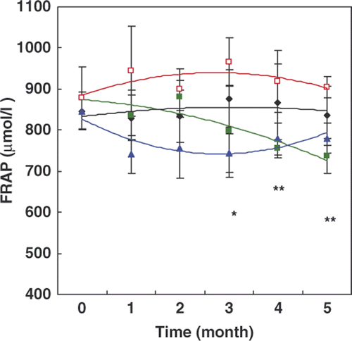
Table II. Comparison of serum Hsp70 levels in control and diabetic rats with or without hot-tub therapy (HTT).
Figure 4. Activity of native (•, black) and denatured Firefly luciferase by GnHCl, in the absence (♦, green) or presence (▪, blue) of native and glycated Hsp70 (▴, red) at different time intervals. The activity of native enzyme (luciferase) was compared to the original activity and its activity in the other samples was compared to the maximum recovery by normal Hsp70 at 20 min.
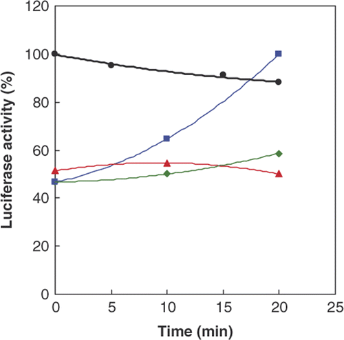
Figure 5. Plot of rats survival (cumulative survival) analysed by the Kaplan-Meier method on all groups of rats. All animals in the control groups with or without HTT (dashed line) survived up to the end of experiment. The number of rats surviving up to the end in diabetic groups were 5 and 3 from 8 (in each group) with (dotted line) or without HTT (continuous line), respectively. The results of statistical analysis are explained in the text.
