Figures & data
Table I. Hsp27 and Hsp70 expression model parameters for PC3 and RWPE-1 cells calculated by where H(t, T) represents Hsp27 and Hsp70 expression normalised with respect to actin concentration.
Figure 1. Western blots depicting Hsp27, Hsp60, and Hsp70 and actin levels for (A) PC3 and (B) RWPE-1 cells heated at 44°C for various heating durations, n = 3.

Figure 2. Distribution of Hsp27 (green fluorescence) and Hsp70 (red fluorescence) in PC3 cells (A) unheated (maintained at 37°C in incubator) and (B) heated (T = 44°C for 5 min) and RWPE-1 cells (C) unheated and (D) heated at T = 44°C for 5 min.

Figure 3. Normalised Hsp27/actin, Hsp60/actin, and Hsp70/actin expression ratios as a function of heating time and temperature determined with western blotting and evaluated at 16 h PH, n = 3 for PC3 cells with an average standard deviation in HSP expression measurement of ±0.12 mg mL−1.
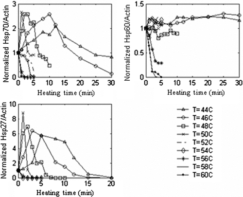
Figure 4. Normalised Hsp27/actin, Hsp60/actin, and Hsp70/actin expression ratios as a function of heating time and temperature determined with western blotting and evaluated at 16 h PH, n = 3 for RWPE-1 cells with an average standard deviation in HSP expression measurement of ±0.14 mg mL−1.
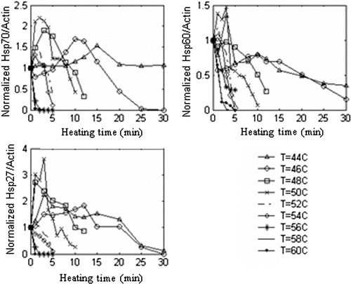
Figure 5. Normalised (A) Hsp27/actin, (B) Hsp60/actin, and (C) Hsp70/actin levels for PC3 cells as a function of heating time at T = 44°C and evaluation at various post-heating (PH) periods with an average standard deviation in HSP expression measurement of ±0.12 mg mL−1, n = 3.
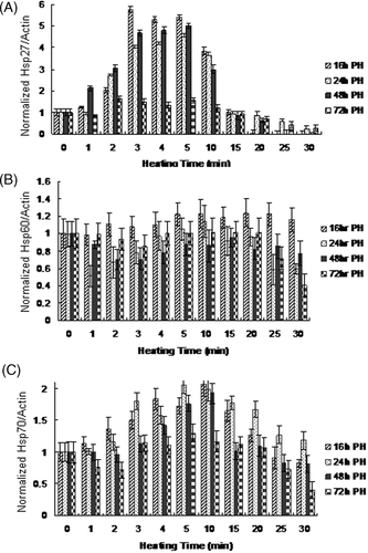
Figure 6. Comparison between measured and model-predicted (A) Hsp27 expression and (B) Hsp70 expression for PC3 cells and (C) Hsp27 expression and (D) Hsp70 expression for RWPE-1 cells.
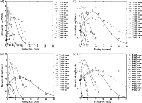
Figure 7. Flow cytometric analysis of cell viability (adapted from Feng et al. Citation[63]). The dead cell population was defined by the marker (M1) as the region of the histogram occupied by both the methanol-treated and severely heat shocked samples (this region excluded the live population defined by the control sample). Cell viability determination is shown for (A) control (unheated), (B) methanol treated, (C) severely heat-shocked (52°C, 6 min), and (D) typical heated sample (44°C, 15 min).
![Figure 7. Flow cytometric analysis of cell viability (adapted from Feng et al. Citation[63]). The dead cell population was defined by the marker (M1) as the region of the histogram occupied by both the methanol-treated and severely heat shocked samples (this region excluded the live population defined by the control sample). Cell viability determination is shown for (A) control (unheated), (B) methanol treated, (C) severely heat-shocked (52°C, 6 min), and (D) typical heated sample (44°C, 15 min).](/cms/asset/55e0da95-8bcb-425b-9300-e2c3f213fe87/ihyt_a_486778_f0007_b.gif)
Figure 8. (A) PC3 cell viability in response to variable thermal stress duration as measured at 72 h PH with the average standard deviation in cell viability measurement of ±3.5%, n = 3, (B) RWPE-1 cell viability in response to variable thermal stress duration as measured at 72 h post heating with the average standard deviation in cell viability measurement of ±2.9%, n = 3.

Figure 9. Comparison of measured cell viability with (A, B) Hsp70 and (C, D) Hsp27 in PC3 cells as a function of temperature and heating duration.
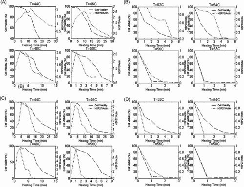
Figure 10. Natural logarithm of time as a function of 1/T for Ω = 1 for (A) PC3 and (B) RWPE-1 cells.
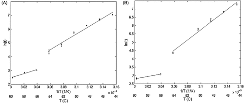
Table II. Values of activation energy, Ea, and frequency factor A, for cell injury calculated by fitting the Arrhenius damage model to measured cell viability data for both cell types.
Figure 11. Comparison between measured and Arrhenius model-predicted damage for (A) PC3 and (B) RWPE-1 cells.

Table III. Minimum threshold heating protocols to diminish Hsp27 and Hsp70 expression below the basal level.