Figures & data
Figure 1. A schematic diagram of the electromagnetic thermal surgery system used in the study. The system was designed in our laboratory and custom-made by a commercial manufacturer.
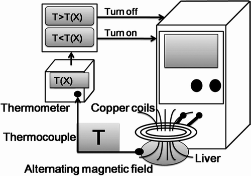
Figure 2. Ex vivo experiments: (A) and (B) temperature measurement by infrared imager. Note that in (A) a single needle was used, while in (B) nine needles were used; (B) produced a much higher temperature than (A). (C) An array of needles was inserted along the resection line over an isolated porcine liver with an interval of 5 mm between each other (arrow). (D) After a 3-min electromagnetic thermocoagulation, the cutting surface of the liver explant showed a completely ‘cooked’ appearance.
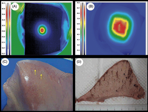
Figure 3. Animal studies in rats (A-C) and rabbits (D-F). Note that needle arrays were aligned along the resection lines (A and D, arrows). After resection, the surfaces of remnant liver were completely coagulated and no bleeding was noted after the operation (B and E). Using electromagnetic thermal therapy, a part of the median lobe of liver was resected successfully (C and F).
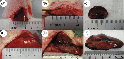
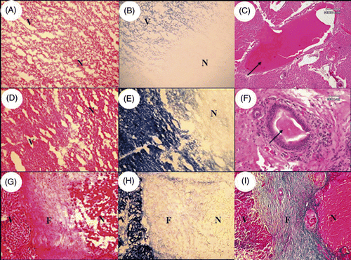
Figure 4. Histological examination of the immediately resected liver (A and B: rat; C-F: rabbit) and the remnant liver after a 30-day recovery (G-I: rabbit). The livers immediately after the resection showed coagulation necrosis with loss of NADPH-diaphorase activity in necrotic area (N) compared to viable liver tissue (V) (A, B, D and E). After the thermocoagulation, the lumen of blood vessel was blocked by thrombus (C, arrow) and the lumen of bile duct was blocked by condensed plugs (F, arrow). After a 30-day recovery, the resected margin of remnant liver showed a fibrous band (F) between viable liver tissue (V) and necrotic tissue (N) indicating a healing process (G-I). (A, D and G: H & E stain, 100×; C: H & E stain, 40×; F: H & E stain, 200×; B, E and H: NADPH-diaphorase stain, 100×; I: Masson-trichrome stain, 100×).