Figures & data
Figure 1. Effect of β-lap alone or with dicoumarol on the clonogenic survival of HOS cells. (A) HOS cells were treated with 5, 7.5 and 10 µM β-lap for 1, 2, 4 and 8 h, cultured for 7–9 days, and the surviving fractions were calculated. Averages of 4 experiments with 1 SE are shown. (B) HOS cells were incubated with 50 µM dicoumarol alone, 7.5 µM β-lap alone or in combination for 1 h at 37°C and the clonogenic survivals were determined. Averages of 4 experiments with 1 SE are shown.
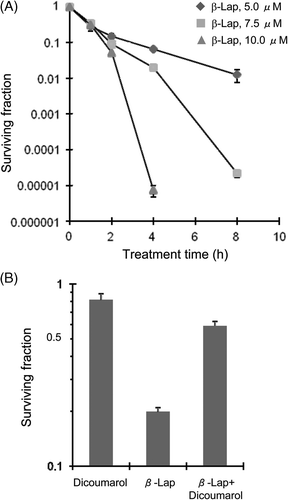
Figure 2. Effect of β-lap on cell cycle and γH2AX expression. HOS cells were treated with 7.5 µM β-lap for 1 h, incubated in regular medium at 37°C, harvested 0, 3, 6, 12, 24, 48, 72 h later, and the cell cycle distribution was analysed with flow cytometry method (A and B). γH2AX formation at different cell cycle phases after treatment with 7.5 µM β-lap for 1 h (C). Cells were treated with 7.5 µM β-lap for 1 h at 37°C, incubated with regular medium for 30 min, immunostained for γH2AX and then the cell cycle distribution was assessed by flow cytometry.
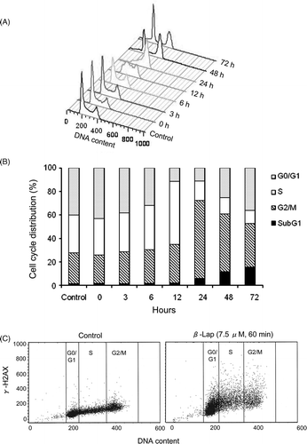
Figure 3. Heat-induced potentiation of the cytotoxicity of β-lap and heat-induced change in the cell cycle distribution. (A) HOS cells were treated with 7.5 µM β-lap for 1 h at different temperatures, washed, cultured for 7–9 days, and the surviving fractions were calculated. Average of 6 experiments is shown. (B) Western blot analysis for NQO1 expression in HOS cells at various times after heating at 42°C for 1 h. (C) Cell survivals were determined after treating the cells with heating at 42°C for 1 h alone, incubation with 7.5 µM β-lap at 37°C for 1 h alone or treating the cells with 7.5 µM β-lap at 42°C for 1 h. Cell survival was also determined after heating the cells at 42°C for 1 h and then treating the cells with 7.5 µM β-lap for 1 h at various times after the heating. The dotted line indicates the expected cell survival when the heating and β-lap treatment reduced the survival of the cells additively. Averages of 6 experiments with 1 SE are shown. (D) Effect of heating on the cell cycle progression is shown. Cells were heated at 42°C for 1 h, incubated in regular medium at 37°C for 0–24 h and the cell cycle distribution was assessed with flow cytometry.
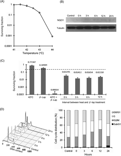
Figure 4. Effect of β-lap and heat treatment on the phosphorylation of H2AX. β-Lap: Cells were incubated with 7.5 µM β-lap for 1h, incubated at 37°C for 30 min and the expression of γH2AX was determined. Heat: cells were heated at 42°C for 1 h, incubated at 37°C for 24 h and the expression of γH2AX was determined. Heat + β-Lap: cells were heated at 42°C for 1 h, incubated at 37°C for 24 h, treated with 7.5 µM β-lap for 1 h, incubated at 37°C for 30 min and the expression of γH2AX was determined. (A) Immunofluorescent staining for γH2AX; (B) average number of γH2AX foci in each cell; (C) percentages of γH2AX positive cells.
