623
Views
14
CrossRef citations to date
0
Altmetric
RESEARCH ARTICLE

KNK437, a benzylidene lactam compound, sensitises prostate cancer cells to the apoptotic effect of hyperthermia

, , &
Pages 63-73 | Received 04 Sep 2009, Accepted 27 Sep 2010, Published online: 04 Jan 2011

Figures & data

Figure 1. (A) Colony numbers of PC-3 cells treated with hyperthermia and KNK437 (100 µM). Cells were preincubated with DMSO (H) or DMSO + KNK437 (KNK437 + H) at 37°C for 5 h before heat shock at 45°C for 1 h. Additionally, cells were preincubated with DMSO (RH) or DMSO + KNK437 (KNK437 + RH) at 37°C for 1 h followed by repeated heating process (first heating at 45°C for 10 min → recovered at 37°C for 4 h → second heating at 45°C for 1 h). Values represent means ± SD of duplicate samples per group; *p < 0.05; **p < 0.01; ***p < 0.001. (B) Comparison of the inhibitory effects of KNK437 (100 µM) and quercetin (100 µM) on the acquisition of thermotolerance in PC-3 cells. Cells were preincubated at 37°C with DMSO (RH), DMSO + KNK437 (KNK437 + RH) and DMSO + quercetin (quercetin + RH) for 1 h followed by repeated heating process as described above. Values represent means ± SD of duplicate samples per group. H, hyperthermia; RH, repeated hyperthermia; *p < 0.05; **p < 0.01; ***p < 0.001.
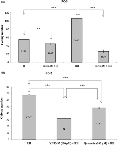
Figure 1. (A) Colony numbers of PC-3 cells treated with hyperthermia and KNK437 (100 µM). Cells were preincubated with DMSO (H) or DMSO + KNK437 (KNK437 + H) at 37°C for 5 h before heat shock at 45°C for 1 h. Additionally, cells were preincubated with DMSO (RH) or DMSO + KNK437 (KNK437 + RH) at 37°C for 1 h followed by repeated heating process (first heating at 45°C for 10 min → recovered at 37°C for 4 h → second heating at 45°C for 1 h). Values represent means ± SD of duplicate samples per group; *p < 0.05; **p < 0.01; ***p < 0.001. (B) Comparison of the inhibitory effects of KNK437 (100 µM) and quercetin (100 µM) on the acquisition of thermotolerance in PC-3 cells. Cells were preincubated at 37°C with DMSO (RH), DMSO + KNK437 (KNK437 + RH) and DMSO + quercetin (quercetin + RH) for 1 h followed by repeated heating process as described above. Values represent means ± SD of duplicate samples per group. H, hyperthermia; RH, repeated hyperthermia; *p < 0.05; **p < 0.01; ***p < 0.001.

Figure 2. Synthesised levels of Hsp70 mRNA of PC-3 (A) and LNCaP (B) cells exposed to hyperthermia with or without KNK437 (100 µM)/quercetin (100 µM). Cells were preincubated with DMSO at 37°C for 150 min in control group (C). Cells were preincubated at 37°C with DMSO (H), DMSO + KNK437 (KNK437 + H), and DMSO + quercetin (quercetin + H) for 60 min and then heated at 43°C for 90 min. After these processes, cells were harvested immediately and total RNA was isolated. The TaqMan PCR reaction was carried out as described in the manufacturer's protocols. Values represent means ± SD of triplicate samples per group; *p < 0.05; **p < 0.01; ***p < 0.001.
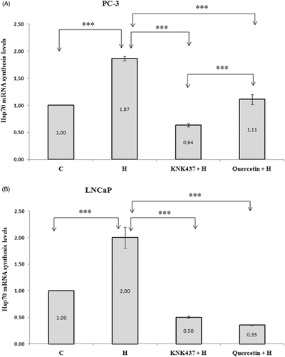
Figure 2. Synthesised levels of Hsp70 mRNA of PC-3 (A) and LNCaP (B) cells exposed to hyperthermia with or without KNK437 (100 µM)/quercetin (100 µM). Cells were preincubated with DMSO at 37°C for 150 min in control group (C). Cells were preincubated at 37°C with DMSO (H), DMSO + KNK437 (KNK437 + H), and DMSO + quercetin (quercetin + H) for 60 min and then heated at 43°C for 90 min. After these processes, cells were harvested immediately and total RNA was isolated. The TaqMan PCR reaction was carried out as described in the manufacturer's protocols. Values represent means ± SD of triplicate samples per group; *p < 0.05; **p < 0.01; ***p < 0.001.
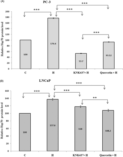
Figure 3. Hsp70 protein levels of PC-3 (A) and LNCaP (B) cells exposed to hyperthermia with or without KNK437 (100 µM)/quercetin (100 µM). Cells were preincubated and treated with drugs and hyperthermia as described in . After treated with drugs and hyperthermia, cells were incubated at 37°C for 24 h and washed with PBS three times, trypsinised and centrifuged. Cell pellet was resuspended with extraction buffer in Hsp70 ELISA kit. Cell suspension was centrifuged at 21,000 g for 10 min at 4°C. Supernatants of samples, 100 µL, were used for analysis of Hsp70 protein levels. The results were expressed as ng/mL. Values represent means ± SD of triplicate samples per group; *p < 0.05; **p < 0.01; ***p < 0.001.

Figure 3. Hsp70 protein levels of PC-3 (A) and LNCaP (B) cells exposed to hyperthermia with or without KNK437 (100 µM)/quercetin (100 µM). Cells were preincubated and treated with drugs and hyperthermia as described in Figure 2. After treated with drugs and hyperthermia, cells were incubated at 37°C for 24 h and washed with PBS three times, trypsinised and centrifuged. Cell pellet was resuspended with extraction buffer in Hsp70 ELISA kit. Cell suspension was centrifuged at 21,000 g for 10 min at 4°C. Supernatants of samples, 100 µL, were used for analysis of Hsp70 protein levels. The results were expressed as ng/mL. Values represent means ± SD of triplicate samples per group; *p < 0.05; **p < 0.01; ***p < 0.001.

Figure 4. Effects of hyperthermia, KNK437 and quercetin on the induction of apoptosis in PC-3 (A) and LNCaP (B) cells. Cells were preincubated and treated with drugs and hyperthermia as described in . Cells treated with hyperthermia and drugs were trypsinised and centrifuged at 1200 g for 5 min at 4°C. An apoptosis detection kit (Biosource International) was used to determine the extent of apoptosis 24 h after treatments. Cell pellet was incubated with annexin-V FITC and PI and incubated at room temperature for 15 min in the dark. Cells were analysed by flow cytometer within 1 h of staining. A minimum of 10,000 cells were collected for each sample. Flow cytometric analysis of the apoptotic cells is shown. The vertical scale represents PI and the horizontal scale represents annexin-V conjugated by FITC. Viable cells, annexin-V-/PI-; dead cells, annexin-V-/PI + ; early apoptotic cells, annexin-V + /PI-; late apoptotic cells, annexin-V + /PI + . Results are representative.
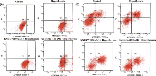
Figure 4. Effects of hyperthermia, KNK437 and quercetin on the induction of apoptosis in PC-3 (A) and LNCaP (B) cells. Cells were preincubated and treated with drugs and hyperthermia as described in Figure 2. Cells treated with hyperthermia and drugs were trypsinised and centrifuged at 1200 g for 5 min at 4°C. An apoptosis detection kit (Biosource International) was used to determine the extent of apoptosis 24 h after treatments. Cell pellet was incubated with annexin-V FITC and PI and incubated at room temperature for 15 min in the dark. Cells were analysed by flow cytometer within 1 h of staining. A minimum of 10,000 cells were collected for each sample. Flow cytometric analysis of the apoptotic cells is shown. The vertical scale represents PI and the horizontal scale represents annexin-V conjugated by FITC. Viable cells, annexin-V-/PI-; dead cells, annexin-V-/PI + ; early apoptotic cells, annexin-V + /PI-; late apoptotic cells, annexin-V + /PI + . Results are representative.

Table I.  Apoptosis index of PC-3 cells exposed to hyperthermia, KNK437 and quercetin.

Table II.  Apoptosis index of LNCaP cells exposed to hyperthermia, KNK437 and quercetin.

Figure 5. The effects of hyperthermia, KNK437 and quercetin on proteolytic cleavage of PARP and activation of caspase-3 in PC-3 (A) and LNCaP (B) cells determined by western blot. Cells were preincubated and treated with drugs and hyperthermia as described in . The cells were washed and disrupted by the addition of lysis buffer containing protease inhibitor cocktail tablet at 4°C and 24 h after hyperthermia and drug treatments of cells. They were then centrifuged at 10,000 g for 30 min at 4°C. Proteins were separated in a 10% (for cleaved-PARP) and 15% (for procaspase-3) SDS-polyacrylamide gel and electroblotted to nitrocellulose membrane. The immunoblotting of cleaved PARP (85 kDa apoptosis related cleavage fragment) and procaspase-3 (32 kDa anticaspase-3 antibody detects inactive form) is shown. Blots are representative. Graph shows the mean ± SD of band intensities (relative arbitrary unit) of three experiments.
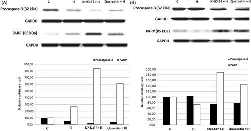
Figure 5. The effects of hyperthermia, KNK437 and quercetin on proteolytic cleavage of PARP and activation of caspase-3 in PC-3 (A) and LNCaP (B) cells determined by western blot. Cells were preincubated and treated with drugs and hyperthermia as described in Figure 2. The cells were washed and disrupted by the addition of lysis buffer containing protease inhibitor cocktail tablet at 4°C and 24 h after hyperthermia and drug treatments of cells. They were then centrifuged at 10,000 g for 30 min at 4°C. Proteins were separated in a 10% (for cleaved-PARP) and 15% (for procaspase-3) SDS-polyacrylamide gel and electroblotted to nitrocellulose membrane. The immunoblotting of cleaved PARP (85 kDa apoptosis related cleavage fragment) and procaspase-3 (32 kDa anticaspase-3 antibody detects inactive form) is shown. Blots are representative. Graph shows the mean ± SD of band intensities (relative arbitrary unit) of three experiments.

Reprints and Corporate Permissions

Please note: Selecting permissions does not provide access to the full text of the article, please see our help page How do I view content?

To request a reprint or corporate permissions for this article, please click on the relevant link below:

Academic Permissions

Please note: Selecting permissions does not provide access to the full text of the article, please see our help page How do I view content?

Obtain permissions instantly via Rightslink by clicking on the button below:

If you are unable to obtain permissions via Rightslink, please complete and submit this Permissions form. For more information, please visit our Permissions help page.