Figures & data
Table I. Keywords used in database searches.
Table II. Summary of thermal damage threshold data compiled in the earlier review updated with newer literature.
Table III. Changes in testicular tubule dimensions in human subjects heated with CEM43 = 180 min.
![Fig 1. Changes in regional blood brain barrier permeability vs. CEM43 after hyperthermia treatment in rats. (A) Fold increase in BBB permeability immediately after whole body heating with a warming pad by Kiyatkin et al. Citation[4]. (B) Increase in BBB permeability immediately following heating in an incubator to CEM43 = 1.3 min by Sharma Citation[5] and 24 h following heating on a warming pad to CEM43 = 0.1–0.7 min by Noor et al. Citation[6]. In (B), some of the data was offset along the x-axis to show all the data points clearly. All data bound by the dashed lines refer to CEM43 = 1.3 min.](/cms/asset/37df625d-1209-4984-9b51-ae0d50bcf0df/ihyt_a_534527_f0001_b.gif)
![Fig 2. Change in cerebral blood flow vs. CEM43. Measurements were made either during or immediately after whole body hyperthermia treatment in rats, humans, and rabbits Citation[5], Citation[7–9].](/cms/asset/6fa0578f-7101-4050-86ed-4ec7e3999bd7/ihyt_a_534527_f0002_b.gif)
![Fig 3. Heat-induced cell death in the rat brain. (A) Cell death in several brain regions at multiple assessment times after whole body hyperthermia treatment in rats at CEM43 = 5.9 min Citation[10]. (B) Fold increase in cell death in several brain regions in rats compared to unheated animals, 10 hours after whole body hyperthermia treatment to CEM43 = 15 min Citation[11].](/cms/asset/29be13c8-c774-4ea2-a4fa-ab3bd5aba60a/ihyt_a_534527_f0003_b.gif)
![Fig 4. Changes in neuronal excitability in two regions of hippocampus following whole body hyperthermia in rats (immediately and 30 min after heating). Data are shown as percent change with respect to baseline measurements (data connected by lines where assessed at both time points). Two different CEM43 are plotted for the mature rats. (A) P1 Amplitude, (B) P1 Onset Latency Citation[15].](/cms/asset/5f2692e7-40d8-4acd-ba6b-81fba0c4c9f3/ihyt_a_534527_f0004_b.gif)
![Fig 5. Heat-induced neurotransmitter concentration changes in the rat brain. (A) glutamate, (B) glycine, and (C) GABA levels are shown in relation to CEM43 in various rat brain regions immediately following whole body hyperthermia Citation[5].](/cms/asset/484257c1-5f46-4d77-8df3-29674c001c90/ihyt_a_534527_f0005_b.gif)
![Fig 6. Changes in rat brain metabolism immediately after whole body hyperthermia. (A) Metabolic changes were assessed by lactate dehydrogenase (LDH) and succinate dehydrogenase (SDH) activity and RNA content at CEM43 = 1.07 min in three rat brain regions Citation[20], and (B) metabolic changes vs. CEM43, as assessed by extracellular brain lactate, pyruvate, glutamate, and glycerol concentrations vs. CEM43 Citation[8].](/cms/asset/83d55f8e-b7b8-4f82-a97c-1f7bf99118a7/ihyt_a_534527_f0006_b.gif)
![Fig 7. Probability of brain lesion induction and blood brain barrier disruption vs. CEM43 in (A) rabbits and rhesus monkeys and (B) pigs Citation[32–36]. In (A), some of the data were offset along the y-axis to show all the data points above CEM43 = 100 min.](/cms/asset/c5740218-cd3a-4142-b414-ce0abda23e25/ihyt_a_534527_f0007_b.gif)
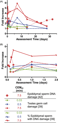
![Fig 8. Decrease in testes weight compared to control. (A) % Decrease in weight of tested vs. assessment time at several thermal doses. (B) Changes in testicular weight assessed at various times, grouped by CEM43. Human data are included for comparative purposes, but human data was reported as a measure of % decrease in volumetric measures. This chart highlights the differences in thermal sensitivity between mouse, rat, and human testes (blue symbols) Citation[37–41].](/cms/asset/51537ad3-3c19-41a0-b70f-698887e73af4/ihyt_a_534527_f0008_b.gif)
![Fig 10. Indications of sperm function after heating in mice and humans. (A) Early effects of heating on sperm characteristics. (B) Late effects of heating on sperm characteristics. Dashed lines: mouse data with a thermal dose of CEM43 = 7.5 min Citation[44]. Solid lines: human data with a thermal dose of CEM43 = 180 min Citation[40].](/cms/asset/fd186d91-dae2-4383-b9ad-c871f9d53e23/ihyt_a_534527_f0010_b.gif)
![Fig 11. Thresholds of thermal damage in eye in rabbits and humans assessed immediately after heating. In rabbits, CEM43 > 21.3 min Citation[73] started causing thermal damage in cornea while thermal lesion in retina was induced at CEM43 = 926 min Citation[75]. In humans, heat treatment at CEM43 = 0.015–34.5 min was not sufficient to cause damage to eyelids Citation[76].](/cms/asset/388c5f09-eeeb-4e46-a508-edfcc772f6c4/ihyt_a_534527_f0011_b.gif)