Figures & data
Figure 1. Experimental planning of the hyperthermia protocols and tumour challenge. (A) Treatment of three groups of mice. CM: administration of intraperitoneal injection of CDDP and DTIC every week. WH: whole body hyperthermia at an interval of 84 h. CO: initial chemotherapy with the same whole body hyperthermia received as WH. (B) Statistically calculated temperature profile of all CO and WH group mice during the third treatment and the infrared thermograph of number 7 and 8 WH mice. Contoured: number 8 WH mouse. (C) Skin temperature of number 8 WH mouse at the end of the third treatment. (D) Inner and surface temperature comparison of number 8 WH mouse during the third treatment.
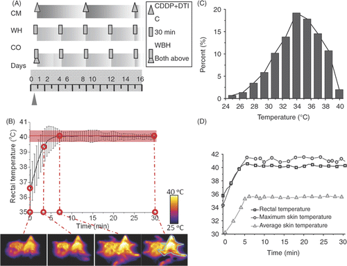
Figure 2. General healthy condition of B16 tumour-bearing mice. (A) The supine appearance of randomly selected mice in each group. Severe depigmentation and alopecia is observed in the drug-treated CM group. The formation of a small pelade in CR group and huge pelade spot in CM group are also distinguishable. Circle: pelade spot. (B) Body weight of C57/BL6 tumour bearing mice after tumour challenge during treatment. Mean ± SD of n = 8 mice/group in CT, CM, WH groups and n = 10/group in CO group. *P < 0.05, **P < 0.01 as compared to the WH group.
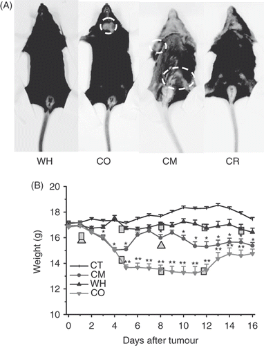
Figure 3. Inhibition of pulmonary metastasis in C57BL/6 mouse model by whole body hyperthermia. The lungs of mice were harvested 16 days after they were inoculated with 3 × 10⁴ cells. (A) Representative macroscopic images of lungs of mice 16 days after intravenous inoculation of B16 melanoma cells and treatment as indicated and photographed under visible light by Cannon camera. (B) Representation of the mean number of metastases (± SD) induced by B16 as indicated in (A). *P < 0.05 with control group.
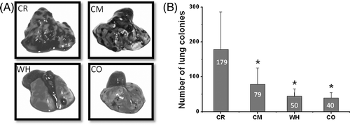
Figure 4. Histological sections of the lungs of mice treated without (A) chemotherapy, (original magnification × 100 and bar 200 µm, local magnification × 200 and bar 100 µm), (B) whole body hyperthermia (magnification × 100, bar 100 µm), (C) combined therapy of hyperthermia and chemotherapy and (D) not treated (magnification × 100, bar 100 µm). Arrowheads: vessel sprout. Circle: metastasis nodules.
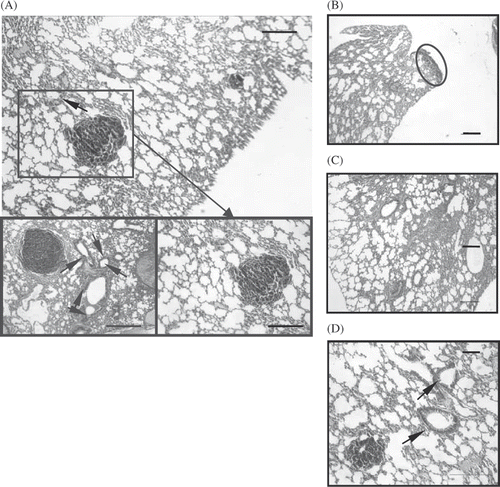
Figure 5. The population of peripheral blood of CD4+ and CD8+ T cells determined by flow cytometry 24 h after the last treatment. (A) Lymphocytes, distinguished by their forward/side light scatter profiles, were analysed by flow cytometry and the percentages of dual-staining positive cells were determined. Results are representative of three independent experiments. (B) CD4+ and CD8+ T cell subpopulations 16 days after tumour challenge indicated treatment; n = 3.
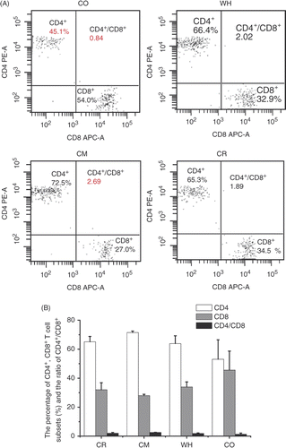
Figure 6. Immunohistochemical determination of ICAM-1, PCNA and cyclin D1 expression in pulmonary metastasis tumours. (A) CO group. The expression of ICAM-1 was strong and that of PCNA and cyclin D1 was weaker; the expression of (B) PCNA and (C) cyclin D1. Both were weak in hyperthermia groups. (D) PCNA expression in the four groups. Magnification: × 400. Bar: 200 µm. All of the stains are positive for PCNA, and the positive cell has a diffuse brown nucleus. In both of the hyperthermia groups, the expression intensity of PCNA in some melanoma cells is weak compared with the control group. In the CR group, numerous cells express the PCNA. All: n = 4. *P < 0.05; **P < 0.1 compared with the WH group.
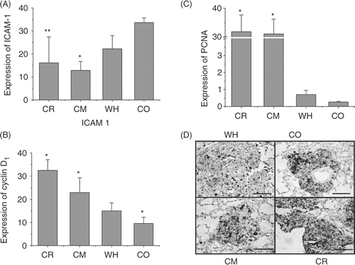
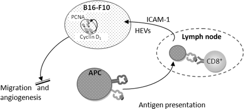
Figure 7. Mechanisms by which whole body hyperthermia inhibits the lung metastasis development of B16-F10.