798
Views
37
CrossRef citations to date
0
Altmetric
Research Article

Contrast-enhanced ultrasound-guided microwave ablation for hepatocellular carcinoma inconspicuous on conventional ultrasound

, , MD, , , &
Pages 555-562 | Received 24 Sep 2010, Accepted 14 Feb 2011, Published online: 28 Jul 2011

Figures & data

Table 1.  Clinical characteristics of patients included (n = 107)

Figure 1. Images in a 64-year-old man who underwent CEUS guidance MW ablation of HCC in segment V of the liver. (A) Conventional US could not detect the tumour. (B) The CEUS arterial phase image shows a 2.2 × 1.4 cm tumour in the right hepatic lobe (arrows). (C) MW antenna was inserted along the guideline under CEUS guidance (arrows). (D) Arterial phase of CEUS obtained 3 months after MW ablation shows complete necrosis of the tumour (arrows).
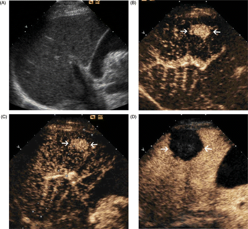
Figure 1. Images in a 64-year-old man who underwent CEUS guidance MW ablation of HCC in segment V of the liver. (A) Conventional US could not detect the tumour. (B) The CEUS arterial phase image shows a 2.2 × 1.4 cm tumour in the right hepatic lobe (arrows). (C) MW antenna was inserted along the guideline under CEUS guidance (arrows). (D) Arterial phase of CEUS obtained 3 months after MW ablation shows complete necrosis of the tumour (arrows).

Figure 2. Images in a 52-year-old male with a 1.8 × 1.5 cm local recurrence of HCC after MW ablation in segment VII of the liver. (A) Contrast-enhanced MRI shows a local recurrence (arrows) in the right hepatic lobe. (B) Conventional US could not detect the tumour. (C) CEUS shows arterial enhancement adjacent to the ablation zone indicative of local recurrence (arrows). (D) MW antenna was inserted along the guideline under CEUS guidance (arrows). (E) Arterial phase of contrast-enhanced MRI obtained 3 months after MW ablation shows complete necrosis of the recurrence (arrows).
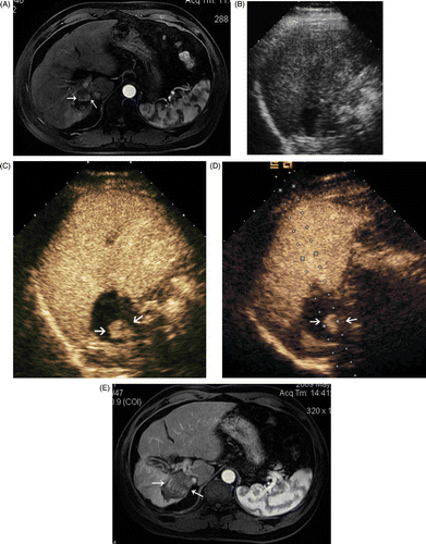
Figure 2. Images in a 52-year-old male with a 1.8 × 1.5 cm local recurrence of HCC after MW ablation in segment VII of the liver. (A) Contrast-enhanced MRI shows a local recurrence (arrows) in the right hepatic lobe. (B) Conventional US could not detect the tumour. (C) CEUS shows arterial enhancement adjacent to the ablation zone indicative of local recurrence (arrows). (D) MW antenna was inserted along the guideline under CEUS guidance (arrows). (E) Arterial phase of contrast-enhanced MRI obtained 3 months after MW ablation shows complete necrosis of the recurrence (arrows).

Reprints and Corporate Permissions

Please note: Selecting permissions does not provide access to the full text of the article, please see our help page How do I view content?

To request a reprint or corporate permissions for this article, please click on the relevant link below:

Academic Permissions

Please note: Selecting permissions does not provide access to the full text of the article, please see our help page How do I view content?

Obtain permissions instantly via Rightslink by clicking on the button below:

If you are unable to obtain permissions via Rightslink, please complete and submit this Permissions form. For more information, please visit our Permissions help page.