Figures & data
Table 1. Effect of regular exercise on embryo survival after maternal hyperthermia, % living embryos/total embryos.
Figure 1. External appearance of ED 8.5 embryos. In the control embryo (C), prosencephalon and diencephalon can be seen through the greatly expanded rhombencephalic roof. Although the roof of its rhombencephalon (RF) is not expanded, the general morphology of the hyperthermia-after-exercise embryo (Ex+H) is similar to that of the control embryo. In addition, distal parts of the neural tube (arrow head) are close to each other. However, the neural tube of the hyperthermia embryo (H) is open (arrow).
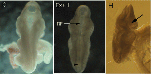
Figure 2. Western blot analysis of Hsp27, Hsp70 and Hsp110 (A). Hsp27 and Hsp110 proteins are highly expressed in the hyperthermia-after-exercise group compared to the hyperthermia group. Densitometric analyses of western blots for Hsp110 (B), Hsp70 (C) and Hsp27 (D) are shown. Data are means ± SEM (n = 32 (C), 30 (E), 18 (Ex + H), 14 (H)). +p < 0.05 comparing the hyperthermia group and the control group of embryos; ++p < 0.001 comparing the hyperthermia group and the control group of embryos; *p < 0.05 comparing the hyperthermia-after-exercise group and the control group. C, control group; E, exercise group; Ex+H, hyperthermia-after-exercise group; H, hyperthermia group.
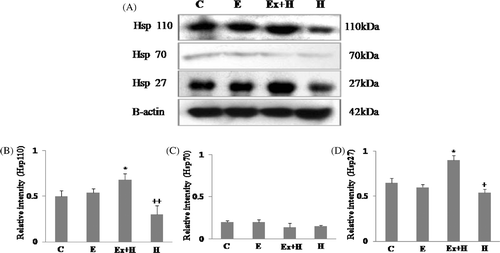
Figure 3. Western blot analysis of Bcl-2 and Bax (A). The hyperthermia-after-exercise group shows significant up-regulation of Bcl-2 protein compared to the hyperthermia group whereas the opposite is true for Bax. Densitometric analyses of western blots of Bcl-2 (B) and Bax (C) are shown. Data are means ± SEM (n as indicated in ). +p < 0.05 comparing the hyperthermia group with the control group; ++p < 0.001 comparing the hyperthermia group with the control group. C, control group; E, exercise group; Ex+H, hyperthermia-after-exercise group; H, hyperthermia group.
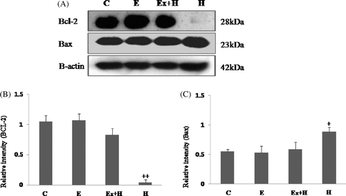
Figure 4. Western blot analysis of cleaved active caspase-9, caspase-7 and caspase-3 (A). Cleaved caspase-9, caspase-7, caspase-3 are up-regulated in the hyperthermia group and down-regulated in the hyperthermia-after-exercise group. Densitometric analyses of western blots of cleaved caspase-9 (B), caspase-7 (C), and caspase-3 (D) are shown. Data are means ± SEM (n as indicated in ). ++p < 0.001 comparing the hyperthermia group and the control group.
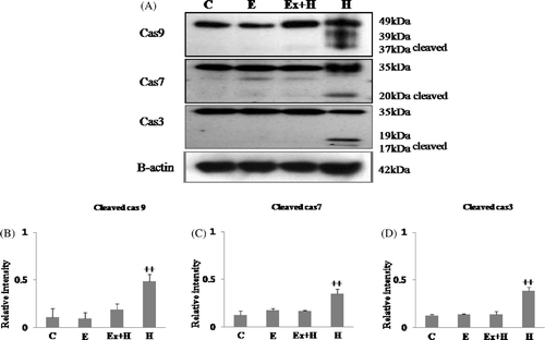
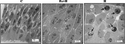
Figure 5. Transmission electron microscopy of the neuroepithelium of prosencephalon. Many more apoptotic cells (arrow) are seen in the hyperthermia group than in the hyperthermia-after-exercise group. C, control group; Ex+H, hyperthermia-after-exercise group; E, exercise group.