Figures & data
Figure 1. The structure of the novel enediyne compounds. (A) PyED; (B) QuinED · ZnCl2.
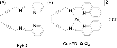
Figure 2. Potentiation of enediyne toxicity by hyperthermia. HeLa CCL-2 cells were treated with various concentrations of either PyED or QuinED · ZnCl2 for 1 h at 37°C or 42.5°C. Surviving fraction was normalised to vehicle only (1.5% DMSO) for each respective temperature. Error bars represent SEM (n = 2 for drug curves and n = 4 for DMSO only curves). (A) PyED; (B) QuinED · ZnCl2.
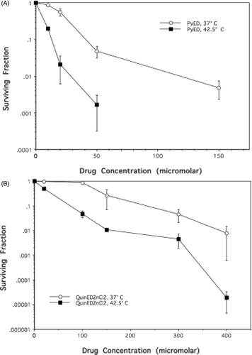
Figure 3. Alterations in the mode of death after treatment with PyED or QuinED · ZnCl2 for 1 h at 37°C or 42.5°C. Apoptosis was measured using the Annexin V/PI assay as described in the Materials and methods section. (A) Bivariate plots of PI (y-axis) versus Annexin V-EFGP (x-axis) fluorescence. HeLa CCL-2 cells were incubated with DMSO or drugs for 1 h at 37°C or 42.5°C for 1 h and fixed 24 h after incubation. Percentage of cells in lower right quadrant was taken as the apoptotic fraction. (B) Apoptotic fractions obtained from bivariate plots were plotted as a function of time for each enediyne treatment. Y-error bars represent SEM (n = 2 for drug curves and n = 4 for DMSO only curves).
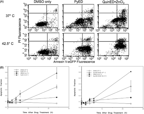
Figure 4. Perturbation of the cell cycle in cells treated with enediynes and hyperthermia. Shown are representative DNA distributions accumulated for HeLa CCL-2 cells incubated with DMSO only or either PyED or QuinED · ZnCl2 for 1 h at 37°C or 42.5°C and fixed 16 h after incubation prior to staining with propidium iodide and analysis by flow cytometry.
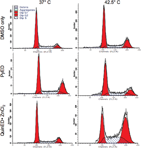
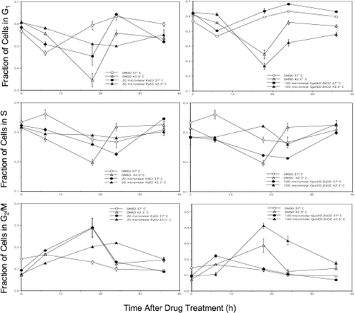
Figure 5. Alterations in cell cycle progression of cells treated with enediynes and hyperthermia. The fraction of cells in each phase of the cell cycle was plotted as a function of time after drug (PyED or QuinED · ZnCl2) and heat treatments. Y-error bars represent SEM (n = 2 for drug curves and n = 4 for DMSO only curves).