Figures & data
Figure 1. Gross pathologic specimens of RF-induced coagulation. Tumours treated with (A) RF alone, (B) liposomal quercetin 24 h pre-RF, (C) liposomal doxorubicin 15 min post-RF, and (D) triple therapy consisting of liposomal quercetin 24 h pre-RF and followed doxorubicin 15 min post-RF are presented. The central white area represents treatment-induced tumour necrosis/coagulation (as noted by the white arrows in each image), with viable tumour staining red. Greater coagulation was observed with RF-quercetin (13.1 ± 0.7 mm) and RF-doxorubicin (13.2 ± 1.3 mm) compared to RF alone (p < 0.001 for both comparisons). The amount of tumour coagulation further increased for triple therapy (quercetin-RF-doxorubicin) (14.5 ± 1.0 mm), compared with quercetin-RF (p = 0.016).
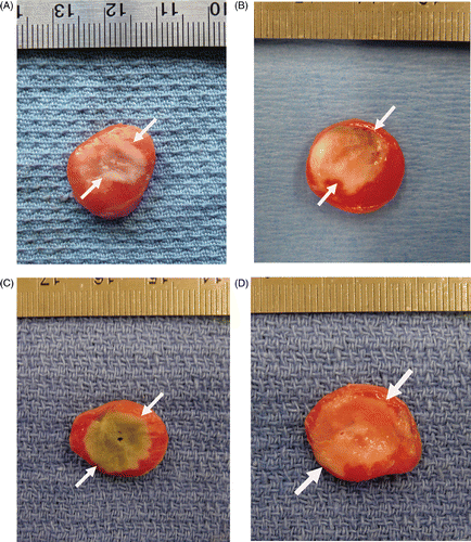
Figure 2. Comparison of tumour coagulation diameter at 4 h and 24 h post-RF ablation in different treatments. The bar graph demonstrates marked differences in coagulation obtained with varied treatment. Greatest coagulation is seen for triple therapy. The amount of tumour coagulation significantly increased from 4 to 24 h for all treatment groups except for quercetin alone (p < 0.001 for all comparisons).
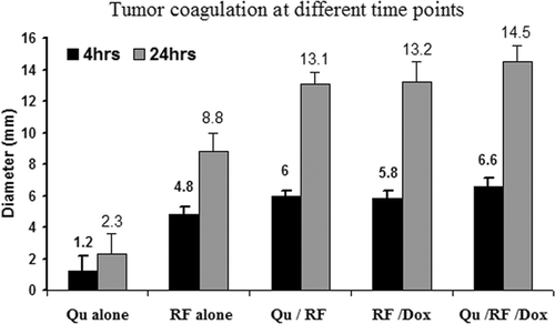
Figure 3. Effect of liposomal quercetin-RF on Hsp70 expression at 4 h. Tumour tissue from quercetin-RF (A, B) stained for Hsp70 demonstrates a weaker band of staining (black arrows) at the periphery of the ablation zone with a lower percentage of positive staining cells compared to RF alone (C, D), noting a marked decrease in heat shock protein formation at 4 h. (A, C = 4×; B, D = 40×).
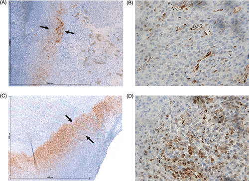
Figure 4. Effect of liposomal quercetin-RF on Hsp70 expression at 24 h. Tumour tissue from quercetin-RF (A, B) stained for Hsp70 demonstrates a comparatively weaker band of staining (black arrows) at the periphery of the ablation zone with a lower percentage of positive staining cells compared to RF alone (C, D), noting a persistent decrease in heat shock protein formation at 24 h. (A, C = 4×; B, D = 40×).
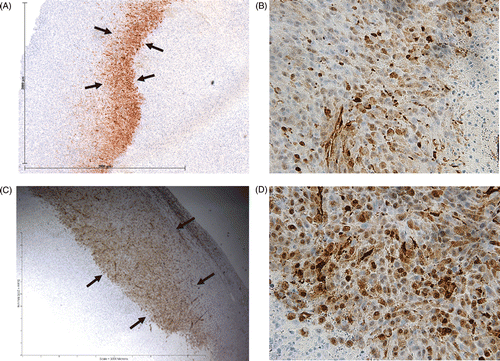
Table 1. Quantitative immunohistochemical staining for heat shock protein expression. There was significantly less Hsp70 expression at the ablative margin for quercetin-RF compared to RF alone at 4 h (rim thickness 0.26 ± 0.08 mm, percentage of stained cells/hpf 22.4 ± 13.9% vs. 0.35 ± 0.18 mm, 38.8 ± 16.1%, P < 0.03). Additionally, RF-doxorubicin resulted in the greatest Hsp70 expression compared to all other groups, including triple therapy (quercetin-RF-doxorubicin) (for both rim thickness and percentage of stained cells/hpf for all comparisons, P < 0.05). Similarly, at 24 h post-treatment, decreased Hsp70 expression was observed for treatments that included quercetin (more so with quercetin-RF than with triple therapy) compared to both RF alone and RF-doxorubicin (for both rim thickness and % stained cells/hpf for all comparisons, P < 0.05).
Figure 5. Effect of liposomal quercetin-RF on apoptosis at 4 h. Tumour tissue from liposomal quercetin-RF (A, B) stained for cleaved caspase-3 demonstrates a thicker band of staining (black arrows) at the periphery of the ablation zone with a greater percentage of positive staining cells compared to RF alone (C, D), noting a marked increase in apoptotic activity at 4 h (A, C = 4×; B, D = 40×).
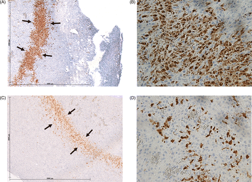
Table 2. Quantitative immunohistochemical staining for apoptosis. Greater cleaved caspase-3 staining was observed for quercetin-RF, RF-doxorubicin, and quercetin-RF-doxorubicin at 4 h, compared to RF alone (for both rim thickness and percentage of stained cells/hpf for all comparisons, P < 0.04 for all comparisons). At 24 h, cleaved caspase-3 expression decreased in all groups (P < 0.001). Quercetin-RF and RF-doxorubicin had comparatively greater cleaved caspase-3 expression than RF alone (for percentage of stained cells/hpf, P < 0.05). Triple therapy with quercetin-RF-doxorubicin had a similar positive cell percentage (45.1 ± 10.8%), to RF-doxorubicin, and was significantly greater compared with other groups (P < 0.03).