Figures & data
Figure 1. Distribution curve of GNPs, based on their particle size (average diameter: 40 nm).
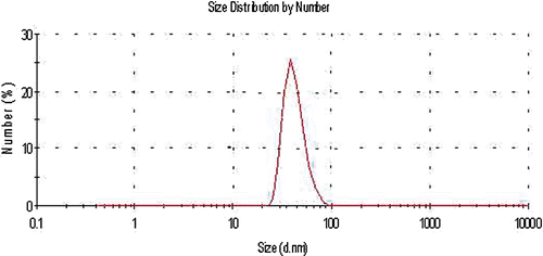
Table I. Treatment conditions of different groups.
Figure 2. Cell survival percentage based on MTT assay 48 h after treatment in the presence and absence of 20 and 40 nm GNPs. Cell incubation time with GNPS was 40 min. The data represent mean ± SD of three performed experiments.
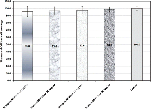
Figure 3. Cell survival percentage based on MTT assay 48 h after hyperthermia in the presence of 20 nm and 40 nm GNPs at 13.2 and 26.4 µg/mL concentrations; also in their absence. Cell incubation time with GNPs was 40 min and MW exposure time was selected as 50 s. The data represent mean ± SD of the three performed experiments.
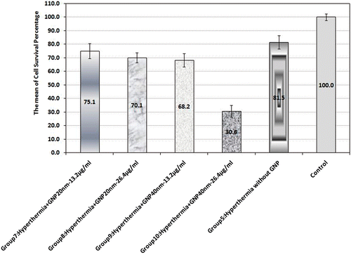
Figure 4. Cell survival percentage based on MTT assay 48 h after hyperthermia, chemotherapy and synergism of both treatments. Cell incubation time with Dox was 30 min and MW exposure time was selected as 50 s. The data represent mean ± SD of the three performed experiments.
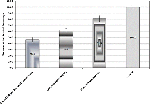
Figure 5. Cell survival percentage based on MTT assay 48 h after chemotherapy in the presence of 20 nm and 40 nm GNPs at 13.2 and 26.4 µg/mL concentrations (groups 12, 13, 14 and 15). Cell incubation times with Dox and GNPs were 30 and 40 min, respectively. The data represent mean ± SD of three performed experiments.
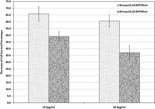
Figure 6. Cell survival percentage based on MTT assay 48 h after combinational therapy (chemotherapy plus hyperthermia) in the presence of 20 nm and 40 nm GNPs at 13.2 and 26.4 µg/mL (groups 16, 17, 18 and 19). Cell incubation times with Dox and GNPs were 30 and 40 min, respectively and MW exposure time was selected as 50 s. The data represent mean ± SD of the three performed experiments.
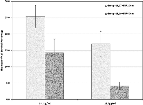
Table II. Relative lethal synergism (RLS) occurred and expected cell death percentage in the presence of 20 nm and 40 nm GNPs. It was calculated as the cell death rate following the use of a therapeutic modality in the presence of GNPs divided by the cell death rate following the same modality but in the absence of GNPs.