Figures & data
Figure 1. Experimental setup.
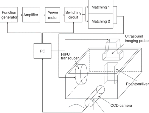
Table I. Specifications of the two HIFU transducers in use.
Table II. Volume ratio (VR) of formed lesions (US/CCD camera) calculated after exposure to US at different electrical powers (and their corresponding acoustic intensities) at either (1) 1 MHz or (2) 3.5 MHz for 60-s, ex-vivo tissue-block experiments.
Figure 2. Real-time monitoring of the lesion-formation process in bovine liver blocks for (A) a 3.5 MHz HIFU transducer at electrical powers of 30, 50, and 70 W. For each power level, B-mode US images were captured after 5, 10, 15, 20, 25, and 30 s of HIFU exposure. The last row shows the optical images of lesions after ablation was completed. Note that no hyperechoes were evident throughout ablation at 30 W. (B) Real-time monitoring of the lesion-formation process in bovine liver blocks for a 1 MHz HIFU transducer at electrical powers of 50, 70, and 90 W. For each power level, B-mode US images were captured after 5, 10, 15, 20, 25, and 30 s of HIFU exposure. Note that no hyperechoes were evident throughout ablation at 50 W. The HIFU transducer was on the left side and the US scanner head was at the top. The ‘+’ represents the focal points of the HIFU transducer. The vertical stripes on most US images, e.g. all images of 3.5 MHz and 30 W, represent the strong acoustic waves from the HIFU transducer picked up by the diagnostic probe.
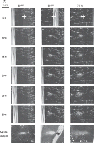
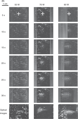
Figure 3. (A) Real-time monitoring of the lesion formation process in transparent tissue-mimicking phantoms for a 3.5 MHz HIFU transducer at electrical powers of 50 and 70 W. For each power level, the upper and lower frames are B-mode US and CCD camera images, respectively, captured after 10, 20, 30, 40, 50, and 60 s of HIFU exposure. The HIFU transducer was on the left side and the US scan head was at the top. (B) Volume ratio (VR) of lesions (US/CCD camera) formed by a 3.5 MHz HIFU transducer for different times and electrical powers. Data are mean and SD values. Dashed line equals 1.0.
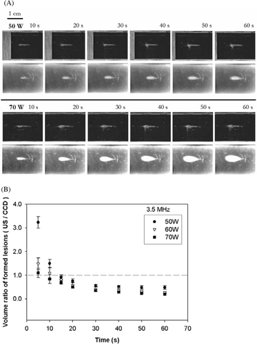
Figure 4. (A) Real-time monitoring of the lesion formation process in transparent tissue-mimicking phantoms for a 1-MHz HIFU transducer at electrical powers of 60 and 70 W. For each power level, the upper and lower frames are B-mode US and CCD camera images, respectively, captured after 10, 20, 30, 40, 50, and 60 s of HIFU exposure. The HIFU transducer was on the left side and the US scanner head was at the top. (B) VR of lesions (US/CCD camera) formed by a 1-MHz HIFU transducer at different times and electrical powers. Data are mean and SD values. Note that there is no value for 60 W before 40 s because no lesion was evident on the CCD camera images. Dashed line equals 1.0.
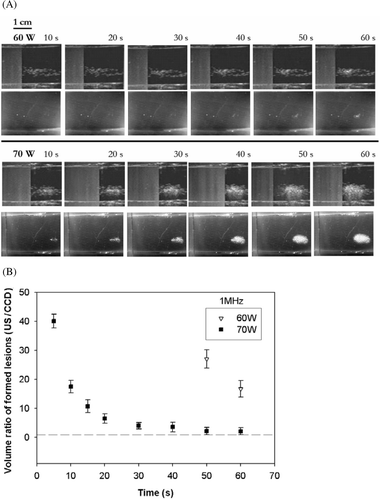
Figure 5. Ultrasonic (left) and optical (right) images of lesions formed by ablations using (A) 1 MHz for 60 s, (B) 3.2 MHz for 60 s, and (C) 1 MHz for 30 s followed by 3.2 MHz for another 60 s (all at 70 W). The ultrasonic image in (C) was shifted left to reveal the few large bubbles to the right of the bubble cloud.
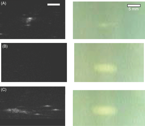
Figure 6. (A) Lesion size and (B) forward shift distance of lesions produced by ablation using 1 MHz for 60 s, 3.2 MHz for 60 s, and 1 MHz for 30 s followed by 3.2 MHz for another 60 s (all at 70 W).
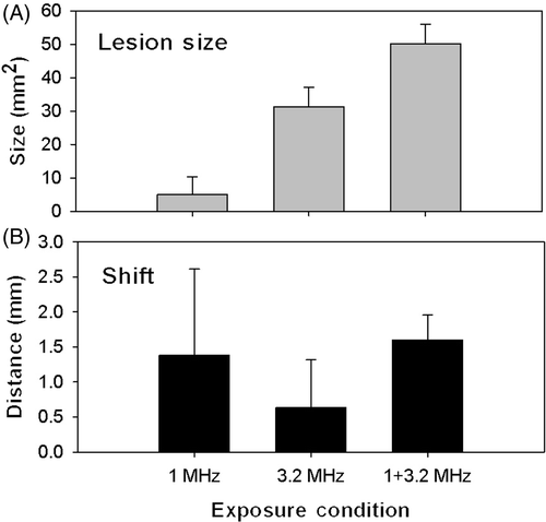
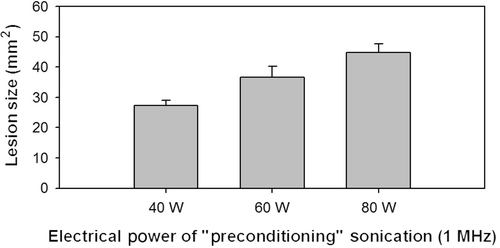
Figure 7. Lesion sizes formed by ablations at 1 MHz for 30 s using electrical powers of either 40, 60 or 80 W followed by 3.2 MHz for 60 s (60 W).