Figures & data
Figure 1. Intracellular ATP in DHD/K12/TRb cells treated with heat only (43° ± 0.05°C for 20 min), Pluronic L10 (A), L61 (B), L64 (C) alone and combined (Pluronic + heat) treatment. *Indicates statistically significant difference (P < 0.05) compared to the untreated control (n = 3 ± STDEV).
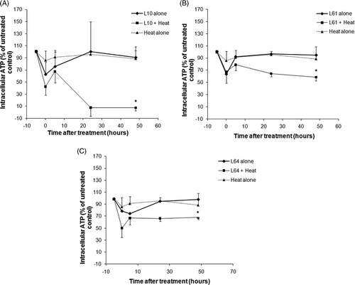
Figure 2. The relative viable cell counts 72 h following treatment with heat (43° ± 0.05°C for 20 min), Pluronic L10, L61, L64 alone and Pluronic + heat. Cell counts were normalised to the untreated control (n = 3). *Indicates significant difference compared to control (P < 0.05).
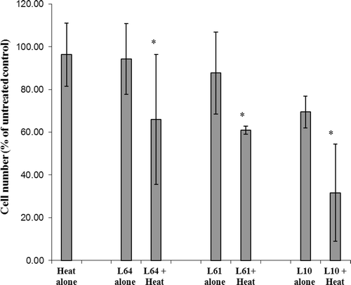
Figure 3. Up-regulation of Hsp70 expression in DHD/K12/TRb cells treated with 43°C low-grade hyperthermia for 20 min (top). Representative western blot of Hsp70 expression with heat stress (bottom). Hsp70 expression at different time points normalised versus actin. Hsp70 expression is elevated 2 h after hyperthermia treatment and lasts 72 h. Each peak is the mean of measurements of 3 ± STDEV.
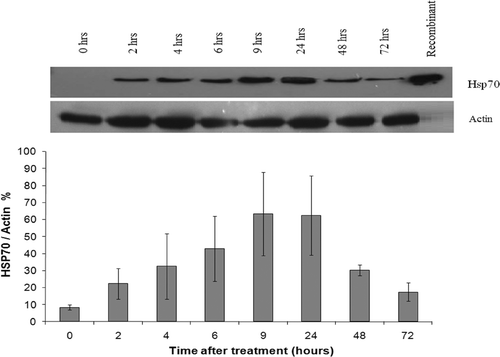
Figure 4. Inhibition of Hsp70 expression in DHD/K12/TRb cells treated with different Pluronics and 43°C low-grade hyperthermia for 20 min. Western blots reveal that the Hsp70 expression was totally diminished by L10 + heat treatment. Pluronic L61, L64, and P85 + heat treatment down-regulate Hsp70 for 6 h.
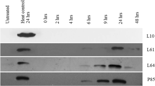
Figure 5. Effect of Pluronic L10, L61, L64, and P85 on the inhibition of Hsp70 with the hyperthermia treatment at 43°C for 20 min. The peaks are normalised to actin and calculated as a percentage relative to heat only treatment at same time points. Results presented as the mean of 3 ± STDEV. *The Hsp70 expression is significantly different from the expression of heat control with P < 0.001.
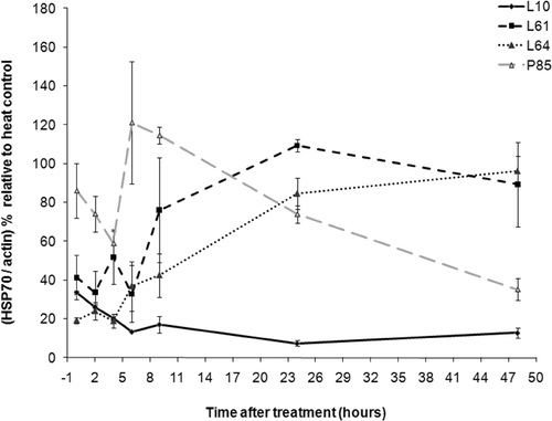
Figure 6. Expression of Hsp70 in DHD/K12/TRb cells treated with Pluronic L10, L61, L64, or P85 for 20 min at 37°C. Western blot analysis indicated an insignificant level of Hsp70 expression with Pluronic only treatment.
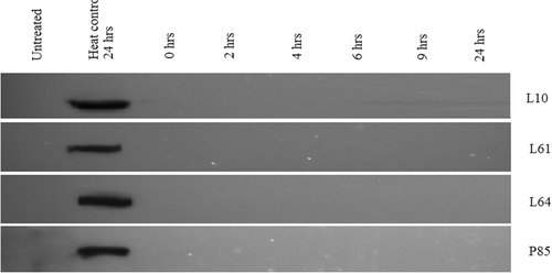
Table I. Summary of Hsp70 expression following treatment with low-grade hyperthermia in conjunction with Pluronic.
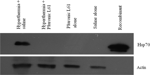
Figure 7. Hsp70 expression 5 h after hyperthermia (for 20 min at 45°C) in subcutaneous colorectal tumour model. A total of 50 µg of tissue lysate was analysed by western blot against anti- Hsp70 antibody. Actin was used as the internal control. Hyperthermia-treated tumours show Hsp70 expression, but with Pluronic L61 the protein expression diminished.