497
Views
19
CrossRef citations to date
0
Altmetric
RESEARCH ARTICLE

Thermal sensitivity of endothelial cells on synthetic vascular graft material

, , , , , PhD & , PhD
Pages 163-174 | Received 31 Oct 2011, Accepted 03 Nov 2011, Published online: 15 Feb 2012

Figures & data

Figure 1 Sketch of the cell seeding process (side view). (A) The 5 mm × 5 mm ePTFE sheet was held in place by a silicone holder's base and gasket (shaded in the diagram) in a standard Petri dish for tissue culture. (B) Cell suspension was pipetted onto the ePTFE sheet. (C) 2.5 mL of cDMEM was added to the dish. Drawings not to scale.
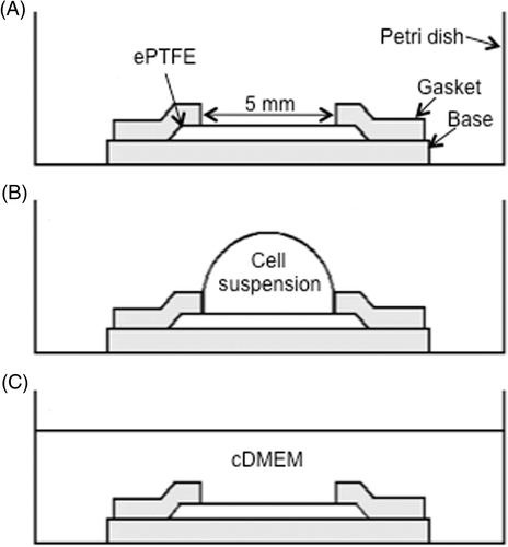
Figure 1 Sketch of the cell seeding process (side view). (A) The 5 mm × 5 mm ePTFE sheet was held in place by a silicone holder's base and gasket (shaded in the diagram) in a standard Petri dish for tissue culture. (B) Cell suspension was pipetted onto the ePTFE sheet. (C) 2.5 mL of cDMEM was added to the dish. Drawings not to scale.

Figure 2 Experimental protocols for detecting immediate or delayed cell death on ePTFE after thermal exposure. Upper left panels, Protocol B: Cells were exposed to various temperatures for various durations, following which the cell viability/death was immediately assessed. Upper right panels, Protocol C: Cells were exposed to various temperatures for 30 min and returned to physiological temperature (37°C) for 20 h. The cell viability/death was then assessed.
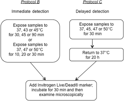
Figure 2 Experimental protocols for detecting immediate or delayed cell death on ePTFE after thermal exposure. Upper left panels, Protocol B: Cells were exposed to various temperatures for various durations, following which the cell viability/death was immediately assessed. Upper right panels, Protocol C: Cells were exposed to various temperatures for 30 min and returned to physiological temperature (37°C) for 20 h. The cell viability/death was then assessed.

Figure 3 Differential survival of heated cells on collagen-coated ePTFE sheets and tissue culture dishes. Cells were cultured at 37°C until they reached confluence on either surface. The percentage of cell death detected after 2 or 4 h at 45°C was almost double on ePTFE sheets compared to tissue culture dishes. The percentage of cell death at 37°C was very low (near zero) on both surfaces.
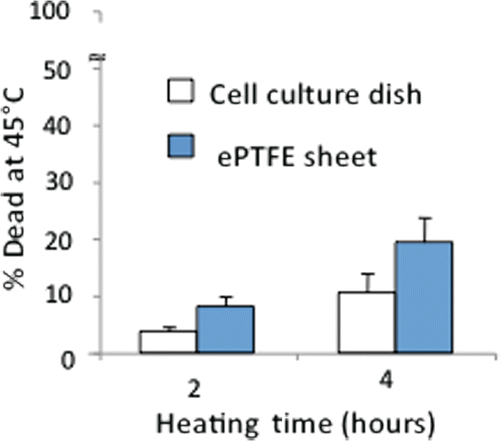
Figure 3 Differential survival of heated cells on collagen-coated ePTFE sheets and tissue culture dishes. Cells were cultured at 37°C until they reached confluence on either surface. The percentage of cell death detected after 2 or 4 h at 45°C was almost double on ePTFE sheets compared to tissue culture dishes. The percentage of cell death at 37°C was very low (near zero) on both surfaces.

Figure 4 Differential adhesion of heated cells onto collagen-coated ePTFE sheets and tissue culture dishes. Cells in suspension were allowed to attach to either surface for 24 h at 37° or 42°C for 24 h. The cells were then stained with calcein AM (which labels the cytosol of viable cells, appearing green) and ethidium homodimer-1 (which labels the nuclei of dead cells, appearing red) and then imaged. At 37°C, endothelial cells formed confluent monolayers and displayed typical cobblestone morphology on both ePTFE sheets (B) and dishes (A). At 42°C, however, the cell density on ePTFE sheets (D) was significantly lower than that on dishes (C). Further, the cells on ePTFE (D) exhibited a condensed cytoplasm, an early indication of apoptosis, though the cells were negative of ethidium homodimer-1 staining.
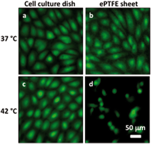
Figure 4 Differential adhesion of heated cells onto collagen-coated ePTFE sheets and tissue culture dishes. Cells in suspension were allowed to attach to either surface for 24 h at 37° or 42°C for 24 h. The cells were then stained with calcein AM (which labels the cytosol of viable cells, appearing green) and ethidium homodimer-1 (which labels the nuclei of dead cells, appearing red) and then imaged. At 37°C, endothelial cells formed confluent monolayers and displayed typical cobblestone morphology on both ePTFE sheets (B) and dishes (A). At 42°C, however, the cell density on ePTFE sheets (D) was significantly lower than that on dishes (C). Further, the cells on ePTFE (D) exhibited a condensed cytoplasm, an early indication of apoptosis, though the cells were negative of ethidium homodimer-1 staining.

Figure 5 Death of cells on ePTFE as determined immediately after various thermal exposures. Cells were cultured on collagen-coated ePTFE at 37°C for a day, and then exposed to 43° and 45°C (A) or 47° and 50°C (B) for various durations. After the heat exposure, the cell viability/death was immediately assessed, and the percentage of dead (necrotic) cells were reported. Significant cell death (more than 50% of cell death) was detected only after long heating durations or exposure at high temperature. Exposure to 37°C served as the control. N = 3–4. Error bars represent SEM. *P < 0.05 compared to the control with the same exposure duration.
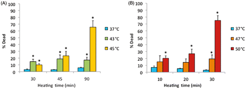
Figure 5 Death of cells on ePTFE as determined immediately after various thermal exposures. Cells were cultured on collagen-coated ePTFE at 37°C for a day, and then exposed to 43° and 45°C (A) or 47° and 50°C (B) for various durations. After the heat exposure, the cell viability/death was immediately assessed, and the percentage of dead (necrotic) cells were reported. Significant cell death (more than 50% of cell death) was detected only after long heating durations or exposure at high temperature. Exposure to 37°C served as the control. N = 3–4. Error bars represent SEM. *P < 0.05 compared to the control with the same exposure duration.

Figure 6 Representative microscopic fluorescent images of subconfluent cells on ePTFE immediately after various thermal exposures. Cells were cultured on collagen-coated ePTFE at 37°C for a day, and then exposed to (A) 37, (B) 45, (C) 47 and (D) 50°C for 30 min. Next, they were immediately stained with calcein AM (which labels the cytosol of viable cells, appearing green) and ethidium homodimer-1 (which labels the nuclei of dead cells, appearing red) and then imaged. Arrows in B–D point to cells that were viable (green) but exhibited a condensed cytoplasm, an early indication of apoptosis, though these cells were negative of ethidium homodimer-1. Most cells at 50°C were necrotic.

Figure 6 Representative microscopic fluorescent images of subconfluent cells on ePTFE immediately after various thermal exposures. Cells were cultured on collagen-coated ePTFE at 37°C for a day, and then exposed to (A) 37, (B) 45, (C) 47 and (D) 50°C for 30 min. Next, they were immediately stained with calcein AM (which labels the cytosol of viable cells, appearing green) and ethidium homodimer-1 (which labels the nuclei of dead cells, appearing red) and then imaged. Arrows in B–D point to cells that were viable (green) but exhibited a condensed cytoplasm, an early indication of apoptosis, though these cells were negative of ethidium homodimer-1. Most cells at 50°C were necrotic.

Figure 7 Cell area on ePTFE as determined immediately after various thermal exposures. Cells were cultured on collagen-coated ePTFE at 37°C for a day, and then exposed to 43° and 45°C (A) or 47° and 50°C (B) for various durations. Next, they were immediately stained with calcein AM (which labels the cytosol of viable cells, appearing green) and ethidium homodimer-1 (which labels the nuclei of dead cells, appearing red), and the area of viable cells was measured and reported as averaged ‘area (µm2)’ per cell. While cell area decreased for all exposures above 37°C, the cell area was the smallest (500 µm2 or less) following the 47° and 50°C/30-min exposures and the 45°C/45-min and 90-min exposures. N = 3–5. Error bars represent SEM. *P < 0.05 compared to 37°C with the same exposure duration. +P < 0.05 compared to the same temperature at the minimum exposure duration.
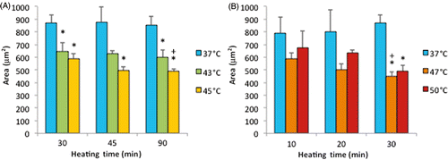
Figure 7 Cell area on ePTFE as determined immediately after various thermal exposures. Cells were cultured on collagen-coated ePTFE at 37°C for a day, and then exposed to 43° and 45°C (A) or 47° and 50°C (B) for various durations. Next, they were immediately stained with calcein AM (which labels the cytosol of viable cells, appearing green) and ethidium homodimer-1 (which labels the nuclei of dead cells, appearing red), and the area of viable cells was measured and reported as averaged ‘area (µm2)’ per cell. While cell area decreased for all exposures above 37°C, the cell area was the smallest (500 µm2 or less) following the 47° and 50°C/30-min exposures and the 45°C/45-min and 90-min exposures. N = 3–5. Error bars represent SEM. *P < 0.05 compared to 37°C with the same exposure duration. +P < 0.05 compared to the same temperature at the minimum exposure duration.

Figure 8 Cell perimeter on ePTFE as determined immediately after various thermal exposures. Cells were cultured on collagen-coated ePTFE at 37°C for a day, and then exposed to 43° and 45°C (A) or 47° and 50°C (B) for various durations. Next, they were immediately stained with calcein AM (which labels the cytosol of viable cells, appearing green) and ethidium homodimer-1 (which labels the nuclei of dead cells, appearing red), and the perimeter of viable cells was measured and reported as averaged ‘perimeter (µm)’ per cell. While cell perimeter decreased for all exposures above 37°C, the cell perimeter was the smallest (100 µm or less) following the 47° and 50°C/30-min exposures and the 45°C/45 and 90-min exposures. N = 3–5. Error bars represent SEM. *P < 0.05 compared to 37°C with the same exposure duration. +P < 0.05 compared to same temperature at the minimum exposure duration.
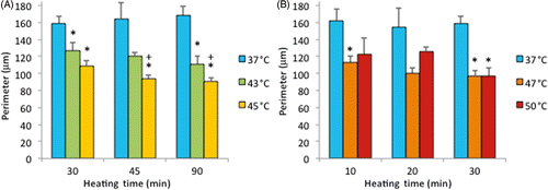
Figure 8 Cell perimeter on ePTFE as determined immediately after various thermal exposures. Cells were cultured on collagen-coated ePTFE at 37°C for a day, and then exposed to 43° and 45°C (A) or 47° and 50°C (B) for various durations. Next, they were immediately stained with calcein AM (which labels the cytosol of viable cells, appearing green) and ethidium homodimer-1 (which labels the nuclei of dead cells, appearing red), and the perimeter of viable cells was measured and reported as averaged ‘perimeter (µm)’ per cell. While cell perimeter decreased for all exposures above 37°C, the cell perimeter was the smallest (100 µm or less) following the 47° and 50°C/30-min exposures and the 45°C/45 and 90-min exposures. N = 3–5. Error bars represent SEM. *P < 0.05 compared to 37°C with the same exposure duration. +P < 0.05 compared to same temperature at the minimum exposure duration.

Figure 9 Cell roundness index on ePTFE as determined immediately following various thermal exposures. Cells were cultured on collagen-coated ePTFE at 37°C for a day, and then exposed to 43° and 45°C (A) or 47° and 50°C (B) for various durations. Next, they were immediately stained with calcein AM (which labels the cytosol of viable cells, appearing green) and ethidium homodimer-1 (which labels the nuclei of dead cells, appearing red), and the roundness index (in the range of 0 and 1) of viable cells was measured and reported as averaged ‘roundness index’ per cell. A perfectly round cell corresponds to an index of 1. Index increased with temperature and exposure duration. N = 3–5. Error bars represent SEM. *P < 0.05 compared to 37°C with the same exposure duration. +P < 0.05 compared to the same temperature at the minimum exposure duration.
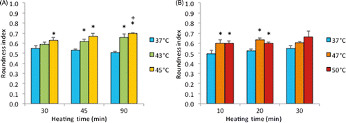
Figure 9 Cell roundness index on ePTFE as determined immediately following various thermal exposures. Cells were cultured on collagen-coated ePTFE at 37°C for a day, and then exposed to 43° and 45°C (A) or 47° and 50°C (B) for various durations. Next, they were immediately stained with calcein AM (which labels the cytosol of viable cells, appearing green) and ethidium homodimer-1 (which labels the nuclei of dead cells, appearing red), and the roundness index (in the range of 0 and 1) of viable cells was measured and reported as averaged ‘roundness index’ per cell. A perfectly round cell corresponds to an index of 1. Index increased with temperature and exposure duration. N = 3–5. Error bars represent SEM. *P < 0.05 compared to 37°C with the same exposure duration. +P < 0.05 compared to the same temperature at the minimum exposure duration.

Table I.  Cell morphology and death on ePTFE immediately after 30-min thermal exposures. Cells were cultured on collagen-coated ePTFE at 37°C for a day, and then exposed to 37° (control), 45°, 47° and 50°C for 30 min. Next, they were stained with calcein AM (which labels the cytosol of viable cells, appearing green) and ethidium homodimer-1 (which labels the nuclei of dead cells, appearing red) for determining the morphology of viable cells and the percentage of dead cells. In general, as the temperature increased, the size (area and perimeter) of viable cells decreased and the percentage of dead cells increased. *P < 0.05 with respect to the 37°C/30-min exposure in the same column.

Figure 10 Delayed cell death on ePTFE upon various thermal exposures. Percentage of cell death was determined following one exposure for 30 min at 37°, 45°, 47° or 50°C with a subsequent 0-h (control) or 20-h incubation period at 37°C. The additional incubation period at 37°C allowed for the progression of apoptosis prior to staining. N = 3–4. Error bars represent SEM. *P < 0.05, compared to the control (zero incubation time after heating) at the same temperature.
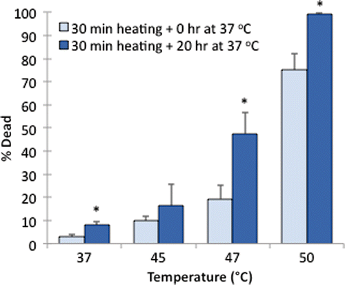
Figure 10 Delayed cell death on ePTFE upon various thermal exposures. Percentage of cell death was determined following one exposure for 30 min at 37°, 45°, 47° or 50°C with a subsequent 0-h (control) or 20-h incubation period at 37°C. The additional incubation period at 37°C allowed for the progression of apoptosis prior to staining. N = 3–4. Error bars represent SEM. *P < 0.05, compared to the control (zero incubation time after heating) at the same temperature.

Figure 11 Representative microscopic fluorescent images of subconfluent cells on ePTFE after various thermal exposures and a delay. Cells were cultured on collagen-coated ePTFE at 37°C for a day, and then exposed to (A) 37°, (B) 45°, (C) 47° and (D) 50°C for 30 min, followed by a 20-h incubation at 37°C that allowed for apoptosis to progress. Next, they were stained with calcein AM (that labels the cytosol of viable cells, appearing green) and ethidium homodimer-1 (that labels the nuclei of dead cells, appearing red) and then imaged. When compared to cells of the same high temperature exposure in , especially the 47° and 50°C exposures (Figures 6C and 6D, respectively), there is an increase in the number of dead cells because the 37°C/24-h incubation allowed for the progression of apoptosis.

Figure 11 Representative microscopic fluorescent images of subconfluent cells on ePTFE after various thermal exposures and a delay. Cells were cultured on collagen-coated ePTFE at 37°C for a day, and then exposed to (A) 37°, (B) 45°, (C) 47° and (D) 50°C for 30 min, followed by a 20-h incubation at 37°C that allowed for apoptosis to progress. Next, they were stained with calcein AM (that labels the cytosol of viable cells, appearing green) and ethidium homodimer-1 (that labels the nuclei of dead cells, appearing red) and then imaged. When compared to cells of the same high temperature exposure in Figure 6, especially the 47° and 50°C exposures (Figures 6C and 6D, respectively), there is an increase in the number of dead cells because the 37°C/24-h incubation allowed for the progression of apoptosis.

Reprints and Corporate Permissions

Please note: Selecting permissions does not provide access to the full text of the article, please see our help page How do I view content?

To request a reprint or corporate permissions for this article, please click on the relevant link below:

Academic Permissions

Please note: Selecting permissions does not provide access to the full text of the article, please see our help page How do I view content?

Obtain permissions instantly via Rightslink by clicking on the button below:

If you are unable to obtain permissions via Rightslink, please complete and submit this Permissions form. For more information, please visit our Permissions help page.