2,357
Views
38
CrossRef citations to date
0
Altmetric
Research Articles

Therapeutic applications of ultrasound in ophthalmology

& , PhD
Pages 405-418 | Received 15 Nov 2011, Accepted 07 Feb 2012, Published online: 23 May 2012

Figures & data

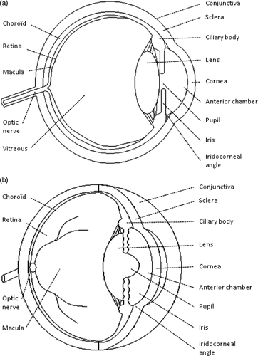
Figure 1. Cross-section (a) and two-point (b) perspective of a human eye.

Figure 1. Cross-section (a) and two-point (b) perspective of a human eye.

Figure 2. Geometry of the six transducers: 4.5-mm width and 7-mm length segment of a 10.2 mm radius cylinder. The dotted lines represent the focal volume, similar to an elliptic cylinder.
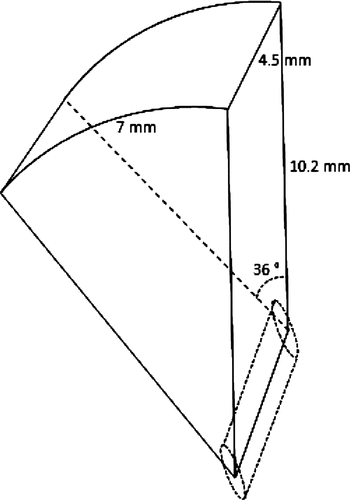
Figure 2. Geometry of the six transducers: 4.5-mm width and 7-mm length segment of a 10.2 mm radius cylinder. The dotted lines represent the focal volume, similar to an elliptic cylinder.

Figure 3. Positioning of the device including circular piezoelectric transducers, inserted into a spacing cone in contact with the eye (A). Sequential activation of the transducers, producing an ultrasonic beam which selectively destroys a well-defined portion of the ciliary body (B). CC: coupling cone. CB: ciliary body. UB: ultrasound beam.

Figure 3. Positioning of the device including circular piezoelectric transducers, inserted into a spacing cone in contact with the eye (A). Sequential activation of the transducers, producing an ultrasonic beam which selectively destroys a well-defined portion of the ciliary body (B). CC: coupling cone. CB: ciliary body. UB: ultrasound beam.

Figure 4. Histological sections at low (top) and high (low) magnification showing the difference between the intact (A and C) and treated (B and D) ciliary body: loss of the bilayered epithelium responsible for the production of aqueous humor (fluid filling the eye).
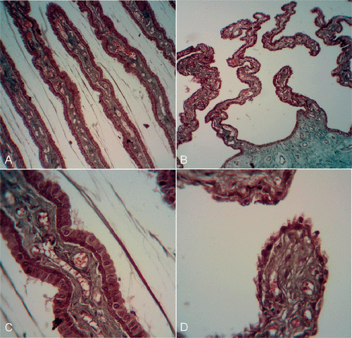
Figure 4. Histological sections at low (top) and high (low) magnification showing the difference between the intact (A and C) and treated (B and D) ciliary body: loss of the bilayered epithelium responsible for the production of aqueous humor (fluid filling the eye).

Table 1.  Summary of the experimental studies evaluating the potential of ultrasound for ocular drug, protein or gene delivery.
Reprints and Corporate Permissions

Please note: Selecting permissions does not provide access to the full text of the article, please see our help page How do I view content?

To request a reprint or corporate permissions for this article, please click on the relevant link below:

Academic Permissions

Please note: Selecting permissions does not provide access to the full text of the article, please see our help page How do I view content?

Obtain permissions instantly via Rightslink by clicking on the button below:

If you are unable to obtain permissions via Rightslink, please complete and submit this Permissions form. For more information, please visit our Permissions help page.