Figures & data
Figure 1. Schematic representation of a polymeric micelle.
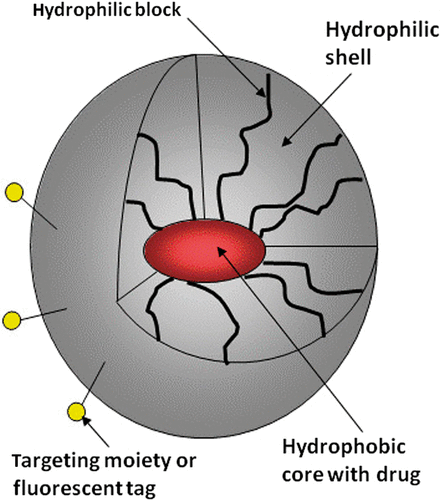
Figure 2. DOX release profiles from 10% poly(ethylene glycol)-co-poly(propylene glycol)-co-poly(ethylene glycol) triblock copolymer (Pluronic) micelles under continuous wave (CW) or pulsed 20 kHz ultrasound at various parameters of ultrasound pulses. Measurements are based on quenching of DOX fluorescence at collisions with water molecules due to the ultrasound-induced DOX transfer from the hydrophobic environment of micelle cores to the aqueous environment. DOX baseline fluorescence is restored between the pulses or when ultrasound is turned off, indicating DOX re-encapsulation in micelle cores. Reprinted from Husseini et al. Citation[85] with permission from ©Elsevier.
![Figure 2. DOX release profiles from 10% poly(ethylene glycol)-co-poly(propylene glycol)-co-poly(ethylene glycol) triblock copolymer (Pluronic) micelles under continuous wave (CW) or pulsed 20 kHz ultrasound at various parameters of ultrasound pulses. Measurements are based on quenching of DOX fluorescence at collisions with water molecules due to the ultrasound-induced DOX transfer from the hydrophobic environment of micelle cores to the aqueous environment. DOX baseline fluorescence is restored between the pulses or when ultrasound is turned off, indicating DOX re-encapsulation in micelle cores. Reprinted from Husseini et al. Citation[85] with permission from ©Elsevier.](/cms/asset/9d4b3bd2-0149-4c2a-82cc-44c67dd37034/ihyt_a_665567_f0002_b.gif)
Figure 3. Effect of Pluronic P-105 micelles on subcellular trafficking of the anthracyclin drug Ruboxyl (Rb) in the multidrug-resistant ovarian carcinoma cells. (A) Rb delivered in PBS does not penetrate into cell nuclei (indicated by arrow). (B) Rb delivered in 1% Pluronic P-105 micelles effectively accumulates in cell nuclei. Reprinted from Rapoport et al. Citation[92] with permission from ©Wiley.
![Figure 3. Effect of Pluronic P-105 micelles on subcellular trafficking of the anthracyclin drug Ruboxyl (Rb) in the multidrug-resistant ovarian carcinoma cells. (A) Rb delivered in PBS does not penetrate into cell nuclei (indicated by arrow). (B) Rb delivered in 1% Pluronic P-105 micelles effectively accumulates in cell nuclei. Reprinted from Rapoport et al. Citation[92] with permission from ©Wiley.](/cms/asset/96870664-80e1-4de0-b634-2f3315c63fac/ihyt_a_665567_f0003_b.gif)
Figure 4. Fluorescence images of A2780 cells. (A) Cells were incubated with Hoechst 33258 to reveal the location of cell nuclei. (B) Cells were incubated with a DOX hydrochloride dissolved in the culture medium; DOX accumulated in cell nuclei. (C) Cells were incubated with hydrophobised DOX encapsulated in PEG-PCL micelles; DOX was internalised by the cells but did not penetrate into cell nuclei. (D) A confocal image of cells sonicated in the presence of DOX encapsulated in PEG-PCL micelles; ultrasound enhanced DOX penetration into cell nuclei but the effect was not uniform. Some small fraction of cells was pre-incubated with Hoechst 33258; Hoechst fluorescence colour was artificially changed to green in order to reveal DOX penetration into cell nuclei by the generation of yellow colour. Adapted from Mohan and Rapoport Citation[99] with permission from ACS Publishing.
![Figure 4. Fluorescence images of A2780 cells. (A) Cells were incubated with Hoechst 33258 to reveal the location of cell nuclei. (B) Cells were incubated with a DOX hydrochloride dissolved in the culture medium; DOX accumulated in cell nuclei. (C) Cells were incubated with hydrophobised DOX encapsulated in PEG-PCL micelles; DOX was internalised by the cells but did not penetrate into cell nuclei. (D) A confocal image of cells sonicated in the presence of DOX encapsulated in PEG-PCL micelles; ultrasound enhanced DOX penetration into cell nuclei but the effect was not uniform. Some small fraction of cells was pre-incubated with Hoechst 33258; Hoechst fluorescence colour was artificially changed to green in order to reveal DOX penetration into cell nuclei by the generation of yellow colour. Adapted from Mohan and Rapoport Citation[99] with permission from ACS Publishing.](/cms/asset/0873f10f-1e62-4568-a2e2-19b51d780198/ihyt_a_665567_f0004_b.gif)
Figure 5. Fluorescence micrographs of tumour, kidney and heart cells; fluorescently labelled stabilised 5% Pluronic micelles were injected intravenously into the A2780 ovarian carcinoma bearing mouse; 4 h after the injection, tumour was sonicated for 30 s by 1 MHz ultrasound at a 3.4 W/cm2 power density. The insert is a confocal image of the cultured tumour cells incubated with a 50 mg/mL fluorescently labelled Pluronic P-105 solution showing that Pluronic molecules were localised in cell membranes and cytoplasmic vesicles but did not penetrate into the nuclei of ovarian carcinoma cells. Adapted from Gao et al. Citation[82] with permission from ACS Publishing.
![Figure 5. Fluorescence micrographs of tumour, kidney and heart cells; fluorescently labelled stabilised 5% Pluronic micelles were injected intravenously into the A2780 ovarian carcinoma bearing mouse; 4 h after the injection, tumour was sonicated for 30 s by 1 MHz ultrasound at a 3.4 W/cm2 power density. The insert is a confocal image of the cultured tumour cells incubated with a 50 mg/mL fluorescently labelled Pluronic P-105 solution showing that Pluronic molecules were localised in cell membranes and cytoplasmic vesicles but did not penetrate into the nuclei of ovarian carcinoma cells. Adapted from Gao et al. Citation[82] with permission from ACS Publishing.](/cms/asset/bf24bba8-d62a-46fc-8fa0-9d5e95a5dad5/ihyt_a_665567_f0005_b.gif)
Figure 6. (A) Fluorescence histograms of tumour cells in (1) unsonicated and (2) sonicated mouse; 5% stabilised Pluronic micelles comprising 0.1% fluorescently labelled Pluronic P-105 were injected intravenously into two A2780 ovarian carcinoma bearing mice; 4 h after the injections, the tumour of one mouse was sonicated twice for 30 s by 1 MHz ultrasound at 3.4 W/cm2 nominal power density at 50% duty cycle; both mice were sacrificed 10 min after sonication. Adapted from Gao et al. Citation[82] with permission from ACS Publishing. (B) Fluorescence histograms of the tumour cells in (1) non-sonicated and (2) sonicated mouse injected with DOX encapsulated in the stabilised Pluronic P-105 micelles. DOX dose was 6 mg/kg; tumour was sonicated for 30 s by 1-MHz CW ultrasound at a power density of 1.7 W/cm2; ultrasound was applied 8 h after the drug injection. Adapted from Gao et al. Citation[82] with permission from ACS Publishing.
![Figure 6. (A) Fluorescence histograms of tumour cells in (1) unsonicated and (2) sonicated mouse; 5% stabilised Pluronic micelles comprising 0.1% fluorescently labelled Pluronic P-105 were injected intravenously into two A2780 ovarian carcinoma bearing mice; 4 h after the injections, the tumour of one mouse was sonicated twice for 30 s by 1 MHz ultrasound at 3.4 W/cm2 nominal power density at 50% duty cycle; both mice were sacrificed 10 min after sonication. Adapted from Gao et al. Citation[82] with permission from ACS Publishing. (B) Fluorescence histograms of the tumour cells in (1) non-sonicated and (2) sonicated mouse injected with DOX encapsulated in the stabilised Pluronic P-105 micelles. DOX dose was 6 mg/kg; tumour was sonicated for 30 s by 1-MHz CW ultrasound at a power density of 1.7 W/cm2; ultrasound was applied 8 h after the drug injection. Adapted from Gao et al. Citation[82] with permission from ACS Publishing.](/cms/asset/f47fb07f-78e3-409d-bea1-77989322719c/ihyt_a_665567_f0006_b.gif)