1,010
Views
12
CrossRef citations to date
0
Altmetric
Research Article

Bipolar versus multipolar radiofrequency (RF) ablation for the treatment of renal cell carcinoma: Differences in technical and clinical parameters

, , , , , , , , & show all
Pages 21-29 | Received 13 Jul 2012, Accepted 12 Nov 2012, Published online: 11 Jan 2013

Figures & data

Figure 1. Clinical example. Bipolar RF ablation was performed in a T1a renal cell carcinoma. (A, B) Dynamic MRI (T2w coronal image and T1w transverse image) for the diagnosis of an exophytic contrast-enhancing renal cell carcinoma in the lower segment of the left kidney (white arrowheads). (C) CT (transverse image) for documentation of correct positioning of the applicator (active tip length of 30 mm) – note the tip of the applicator was positioned slightly beyond the tumour margin. (D, E, F) Dynamic MRI (T2w coronal image, T1w transverse image and T1w coronal image) 22 months after bipolar RF ablation without evidence of local recurrence – note signs of complete tumour destruction with fat tissue necrosis and lack of suspect contrast-enhancement (white arrowheads).
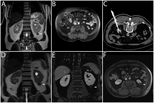
Figure 1. Clinical example. Bipolar RF ablation was performed in a T1a renal cell carcinoma. (A, B) Dynamic MRI (T2w coronal image and T1w transverse image) for the diagnosis of an exophytic contrast-enhancing renal cell carcinoma in the lower segment of the left kidney (white arrowheads). (C) CT (transverse image) for documentation of correct positioning of the applicator (active tip length of 30 mm) – note the tip of the applicator was positioned slightly beyond the tumour margin. (D, E, F) Dynamic MRI (T2w coronal image, T1w transverse image and T1w coronal image) 22 months after bipolar RF ablation without evidence of local recurrence – note signs of complete tumour destruction with fat tissue necrosis and lack of suspect contrast-enhancement (white arrowheads).

Figure 2. Clinical example. Multipolar RF ablation with two applicators was performed in a T1a renal cell carcinoma. (A, B) Dynamic MRI (T2w coronal image and T1w coronal image) for the diagnosis of a renal cell carcinoma with an exophytic tumour in the lower segment of the right kidney showing peripheral nodular contrast-enhancement (white arrowheads). (C) CT (curved maximum intensity projection) for documentation of correct positioning of the applicators (active tip lengths of 40 mm) – note exact parallel positioning of the applicators with a distance of approximately 20 mm. (D) CT (transverse image) directly after multipolar RF ablation – note regular hyperacute appearance of the coagulation with gas bubbles within the treated tumour (white arrowhead). (E, F) Dynamic MRI (T2w coronal image and T1w coronal image) one month after multipolar RF ablation without evidence of local recurrence – note signs of complete tumour destruction without suspect contrast-enhancement (white arrowheads).
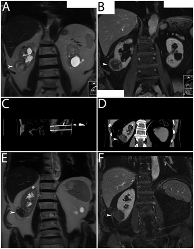
Figure 2. Clinical example. Multipolar RF ablation with two applicators was performed in a T1a renal cell carcinoma. (A, B) Dynamic MRI (T2w coronal image and T1w coronal image) for the diagnosis of a renal cell carcinoma with an exophytic tumour in the lower segment of the right kidney showing peripheral nodular contrast-enhancement (white arrowheads). (C) CT (curved maximum intensity projection) for documentation of correct positioning of the applicators (active tip lengths of 40 mm) – note exact parallel positioning of the applicators with a distance of approximately 20 mm. (D) CT (transverse image) directly after multipolar RF ablation – note regular hyperacute appearance of the coagulation with gas bubbles within the treated tumour (white arrowhead). (E, F) Dynamic MRI (T2w coronal image and T1w coronal image) one month after multipolar RF ablation without evidence of local recurrence – note signs of complete tumour destruction without suspect contrast-enhancement (white arrowheads).

Table I.  Tumour and coagulation extent.

Figure 3. Graphic representation of coagulation extent. (A) Plot of length, width and volume – note there were statistically significant differences between bipolar RF ablation and multipolar RF ablation regarding medians (p < 0.01, p < 0.05 and p < 0.05, respectively). (B) Scatterplot of distance of the applicators versus width for multipolar RF ablation – note dependence of both parameters.
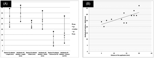
Figure 3. Graphic representation of coagulation extent. (A) Plot of length, width and volume – note there were statistically significant differences between bipolar RF ablation and multipolar RF ablation regarding medians (p < 0.01, p < 0.05 and p < 0.05, respectively). (B) Scatterplot of distance of the applicators versus width for multipolar RF ablation – note dependence of both parameters.

Table II.  Technical data.

Figure 4. Clinical example. Bipolar RF ablation was performed in a T1a renal cell carcinoma. (A) CT (transverse image) for the diagnosis of an intraparenchymal renal cell carcinoma in the upper segment of the right kidney (white arrowhead). (B) CT (transverse image) for documentation of the applicator (active tip length of 20 mm) – note eccentric applicator position in relation to the tumour centre. (C, D) Dynamic CT (transverse images) 19 months after bipolar RF ablation with evidence of local recurrence – note signs of incomplete tumour destruction with suspect contrast-enhancement (white arrowheads).
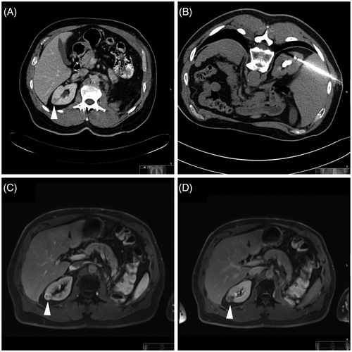
Figure 4. Clinical example. Bipolar RF ablation was performed in a T1a renal cell carcinoma. (A) CT (transverse image) for the diagnosis of an intraparenchymal renal cell carcinoma in the upper segment of the right kidney (white arrowhead). (B) CT (transverse image) for documentation of the applicator (active tip length of 20 mm) – note eccentric applicator position in relation to the tumour centre. (C, D) Dynamic CT (transverse images) 19 months after bipolar RF ablation with evidence of local recurrence – note signs of incomplete tumour destruction with suspect contrast-enhancement (white arrowheads).

Reprints and Corporate Permissions

Please note: Selecting permissions does not provide access to the full text of the article, please see our help page How do I view content?

To request a reprint or corporate permissions for this article, please click on the relevant link below:

Academic Permissions

Please note: Selecting permissions does not provide access to the full text of the article, please see our help page How do I view content?

Obtain permissions instantly via Rightslink by clicking on the button below:

If you are unable to obtain permissions via Rightslink, please complete and submit this Permissions form. For more information, please visit our Permissions help page.