Figures & data
Figure 1. The inhibition effect on OS732 and MG63 cell lines at 43 °C for 1 h measured by MTT. (A) The survival rate of OS732 with different concentration of paclitaxel. (B) The survival rate of OS732 with different concentration of cisplatin. (C) The survival rate of OS732 with combination of 10 μg/mL paclitaxel and 5 μg/mL cisplatin. (D) The survival rate of MG63 with different concentration of paclitaxel. (E) The survival rate of MG63 with different concentration of cisplatin. (F) The survival rate of MG63 with combination of 10 μg/mL paclitaxel and 5 μg/mL cisplatin.
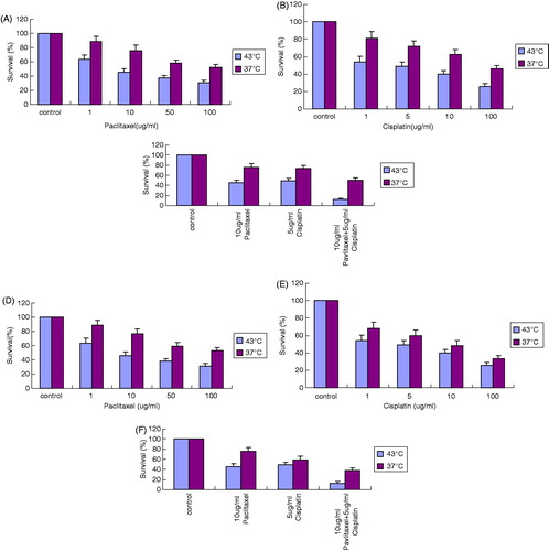
Figure 2. Clonogenic assay of OS cell lines. (A) Clonogenic rate of OS732 cell line. (B) Clonogenic rate of MG63 cell line.
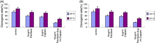
Figure 3. Morphological changes of OS cells with different drugs at 43 °C for 1 h. (A) Morphological appearance of OS cells under inverted phase contrast microscope ×400. (B) Morphological changes of OS cells treated with 10 μg/mL paclitaxel under inverted phase contrast microscope ×400. (C) Morphological changes of OS cells treated with 5 μg/mL cisplatin under inverted phase contrast microscope ×400. (D) Morphological changes of OS cells treated with combination of 10 μg/mL paclitaxel with 5 μg/mL cisplatin under inverted phase contrast microscope ×400. (E) Fluorescent staining of OS cells under the fluorescence microscope ×400. (F) Fluorescent staining of OS cells treated with 10 μg/mL paclitaxel under the fluorescence microscope ×400. (G) Fluorescent staining of OS cells treated with 5 μg/mL cisplatin under the fluorescence microscope ×400. (H) Fluorescent staining of OS cells treated with 10 μg/mL paclitaxel and 5 μg/mL cisplatin under the fluorescence microscope ×400.
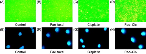
Figure 4. The apoptotic rate of OS cell lines at 43 °C for 1 h measured by FCM. (A) Apoptotic rate of OS732 treated with different concentrations of paclitaxel. (B) Apoptotic rate of OS732 treated with different concentration of cisplatin. (C) Apoptotic rate of OS732 treated with combination of 10 μg/mL paclitaxel and 5 μg/mL cisplatin. (D) Apoptotic rate of MG63 treated with different concentration of paclitaxel. (E) Apoptotic rate of MG63 treated with different concentration of cisplatin. (F) Apoptotic rate of MG63 treated with combination of 10 μg/mL paclitaxel and 5 μg/mL cisplatin.
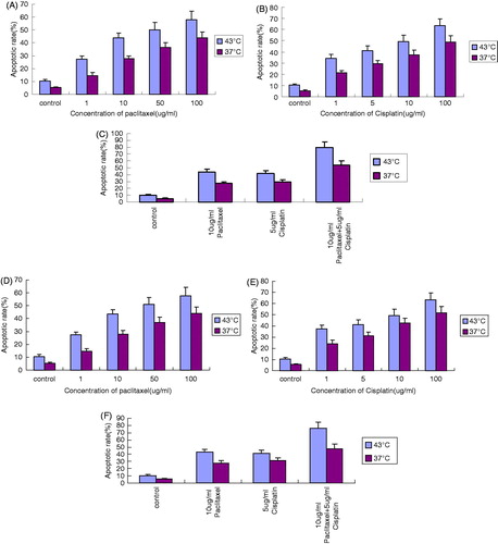
Figure 5. Western blot analysis of Fas. (A) Fas expression of OS cells by WB at 43 °C. (B) Fas expression of OS cells by WB at 37 °C. (C) Quantitative analysis of Fas expression of OS cells.
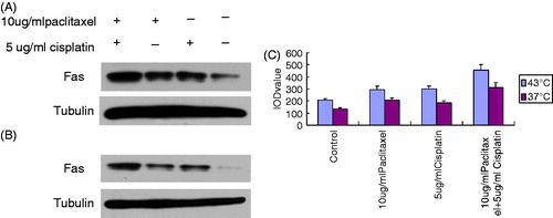
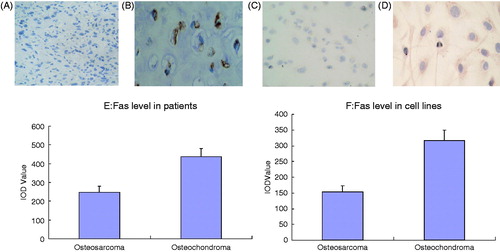
Figure 6. Changes of Fas level of OS in vivo and in vitro. (A) Fas (−) expression of OS patient ×400. (B) Fas (+) expression of osteochondroma patient ×400. (C) Fas (−) expression of OS cells ×400. (D) Fas (+) expression of osteochondroma cells ×400. (E) Quantitative analysis of Fas expression of OS patients and osteochondroma patients. (F) Quantitative analysis of Fas expression of OS cells and osteochondroma cells.