Figures & data
Figure 1. Scheme for the functionalisation procedure of magnetite nanoparticles (MNP) described in this work. (A) Preparation of stabilised magnetite nanoparticle by citric acid (CA). (B) Preparation of cationised albumin by modification of the carboxylate groups with an ethylenediamine. (C) Surface modification of MNP-CA by cationic albumin.
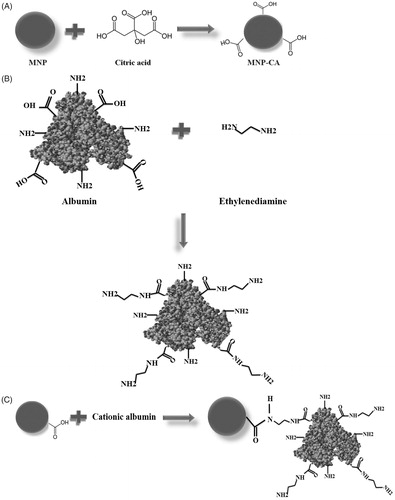
Figure 2. XRD pattern of MNP shows the composition and crystal structure of magnetite.
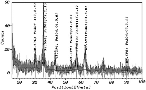
Figure 3. TEM micrograph of MNP-CA.
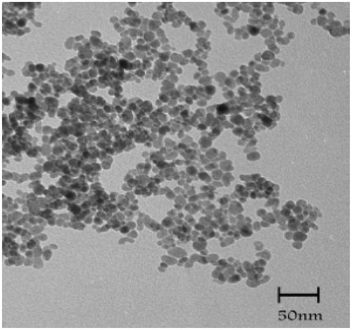
Figure 4. (A) FTIR of citric acid and citrate-capped iron oxide nanoparticles. (B) FTIR of albumin and cationised albumin.
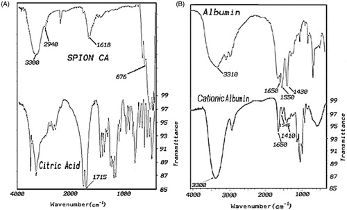
Figure 5. VSM plot of MNP and cationic albumin-conjugated MNP-CA.
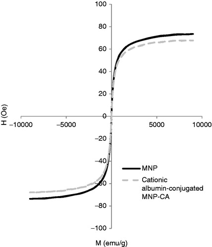
Figure 6. Zeta potential measurements of MNP (A), MNP-CA (B), cationic albumin (C), and cationic albumin-conjugated MNP-CA (D).
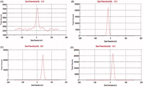
Figure 7. SEM micrograph, zeta size and particle size measurement of cationic albumin-conjugated magnetite nanoparticles.
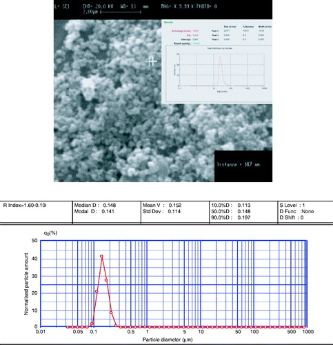
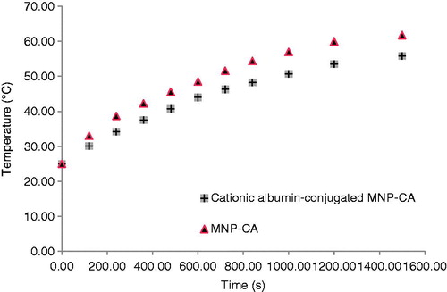
Figure 8. Temperature as a function of time for MNP-CA and cationic albumin-conjugated MNP-CA suspended in distilled water under alternating magnetic field of 215 kHz.