Figures & data
Figure 1. Whole body hyperthermia (WBH) increases the numbers of NK cells and apoptotic cells in colon tumours. (A, B) Localisation of apoptotic cells by TUNEL assay in a patient tumour xenograft from control (A) or WBH treated (B) SCID mice (arrows, original mag. 40X). (C, D) Immunohistochemical localisation of NKp46+ cells in CT26 tumours from control (C) and WBH treated (D) mice at 24 h post-WBH (arrows, original magnification ×10).
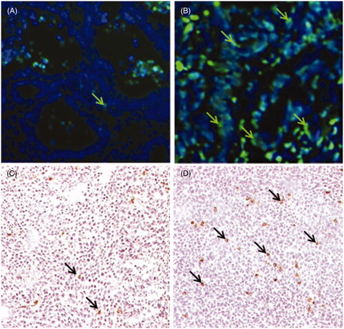
Figure 2. Silencing of (mRNA) HSF1 in tumour cells results in decreased MICA expression and impaired thermally enhanced cytotoxicity by human NK cells. Colo205 cells were transfected with two different siRNA constructs against HSF1 (3134 and 115674) at two different concentrations 3 pg/cell (▪) and 5 pg/cell (□), or with control siRNA at 5 pg/cell ![]()
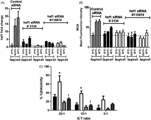
Figure 3. Mutation of HSF1 binding site results in the loss of thermally enhanced activity of MICA promoter. (A) Schematic representation of plasmid constructs containing luciferase reporter gene driven by MICA promoter. Basic pGL3 construct (pGL3), pGL3 construct carrying the MICA promoter (pGL3.MICA), point mutations in HSRE in MICA promoter (pGL3.HSRE), and positive control construct carrying an SV40 promoter and enhancer (pGL3.SV40). (B) Colo205 cells were transfected with the reporter constructs described in (A) and treated at normothermic (▪) or hyperthermic (□) temperatures for 6 h. Relative luciferase activity was detected by dual luciferase reporter assay and normalised to Renilla luciferase activity as transfection control. *p ≤ 0.05 compared to the corresponding normothermic group.
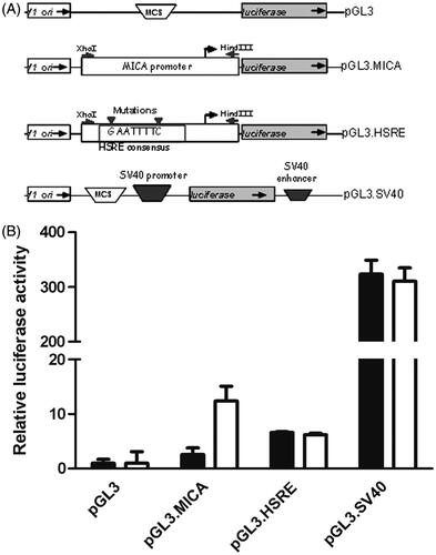
Figure 4. Cycloheximide inhibits induction of MICA by thermal stress in Colo205 cells. Colo205 cells were treated with mild thermal stress at 39.5 °C (▴, ▾) or at 37 °C (♦, •) with DMSO for 6 h in vitro, followed by cycloheximide (5 μg/mL in the culture medium) (▾, •) or DMSO, the solvent used for cycloheximide (▴, ♦) at normothermic temperatures. Cell surface MICA expression was detected with flow cytometry.
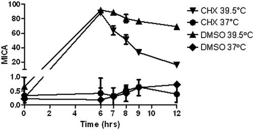
Figure 5. Response of colon cell lines and patient tumour xenografts to mild thermal stress. Colo205 (A), or HT29 (B) tumour bearing SCID mice were treated with whole body hyperthermia (WBH) at 39.5 °C for 6 h in vivo. Fold change of MICA. HSF1 and HSP70 mRNA in the tumours compared to 18 S RNA or levels in tumours from control mice was measured by quantitative RT-PCR and calculated against 18 S RNA, using triplicate wells. The data shown represent the mean ± SD (n ≥ 3) of three experiments. (C) Colon tumours from four patients were implanted into SCID mice and treated with WBH. MICA mRNA expression was detected by qPCR and fold change determined compared to untreated controls (D, E) Kinetics of MICA mRNA levels in Colo205 and HT29 xenografts respectively at 3, 6, 12 and 24 h after WBH. (F) Kinetics of the murine MICA orthologue, Rae1 mRNA expression in CT26 tumours in BALB/c mice □ hyperthermic, ▪ normothermic.
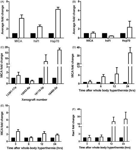
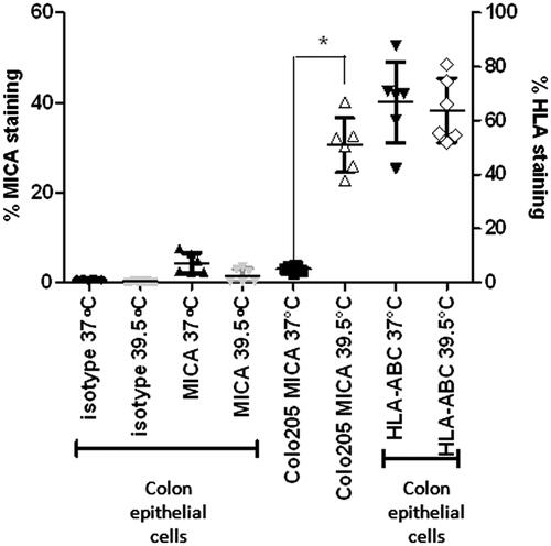
Figure 6. Expression levels of MICA and MHC Class I are not altered with thermal stress in non-malignant, normal human colon epithelial cells. Normal colon epithelial cells isolated from donors and Colo205 cells were treated with mild thermal stress at 39.5 °C for 6 h in vitro and cell surface levels of MICA and HLA-A, B, C were quantified by flow cytometry. The results are plotted showing means and standard deviations of six samples. *p = 0.0002 compared to the corresponding normothermic group using unpaired t-test with Welch’s correction.