Figures & data
Figure 1. Heat-induced radiosensitisation. Cells were heated at 44°C for 15 min (open symbols) or not heated (closed symbols), and then irradiated by X-ray at the indicated doses on the abscissa. Triangles, wild-type cells; circles, DNA-PKcs deficient cells. (A) mouse SC3VA2 (scid, DNA-PKcs−/−) and RD13B2 (hybrid, wild); (B) mouse PK33N (DNA-PKcs) and CB17 (wild); (C) Chinese hamster V3 (DNA-PKcs) and CHO-K1 (wild); (D) human M059J (DNA-PKcs) and M059K (wild).
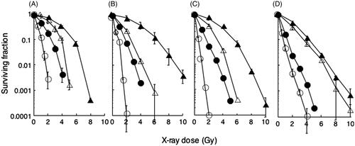
Figure 2. Effect of heat exposures prior to X-ray irradiation on DSB repair. (A) hybrid cells (closed symbols). (B) scid cells (open symbols). Cells were exposed to heat at 44 °C and then irradiated with 20 Gy of X-ray. Circles: non-heat-exposed cells; squares: heat exposure for 15 min; triangles: heat exposure for 30 min; reverse triangles: heat exposure for 45 min. These data are averages of four independent experiments.
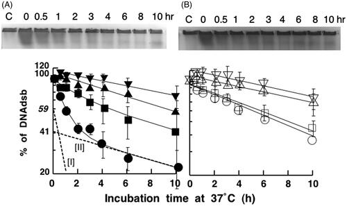
Figure 3. DNA-PK reactivation after a heat exposure in hybrid cells. (A) Hybrid cells were heat-treated at 44 °C for 15 (•), 30 (▴) and 45 (▾) min without irradiation and then cultured at 37 °C for the indicated time on the abscissa. The percentage of DNA-PKcs activity is shown on the vertical axis. (B) The fast repair fraction is indicated on the vertical axis from . The relative DNA-PKcs activity during an incubation from 0 h to 3 h after heat exposure at 44 °C for 15 min (▵) or 30 min (▿) was calculated from results of compared with the non-heated case (○). The abscissa is indicated with a logarithmic scale. Other details are described in the text.
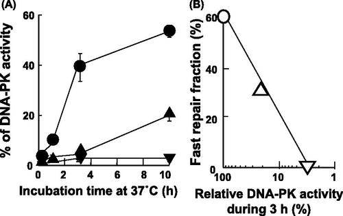
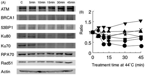
Figure 4. Heat sensitivity of repair proteins from hybrid cells. Results of Western blot (A) and the analysis by densitometry (B) are shown. Key: ▾ ATM; ▪ BRCA1; ★ 53BP1; • Ku70; ♦ Ku80; × RPA70; ▴ Rad51.