Figures & data
Figure 1. Axial T2 MR imaging: mucinous ascites with several peritoneal implants. White arrow shows the mucinous ascites over the liver surface.
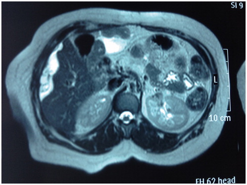
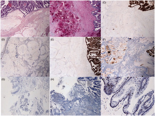
Figure 2. (A) Haematoxylin and eosin-stained section from tumour tissue in the abdominal cavity: there is extensive dissection of mucous material in the adipose tissue and mucinous epithelium with little nuclear atypia (20×). (B) The mucous material is positive with PAS-diastase and (C) MUC4. The epithelium (D) is CK7 weak-irregular positive but (E) CK17 and (F) CEA strongly positive; (G) CK20 and (H) CA125 are negative. (I) p53 shows nuclear positivity in only half of the tumour cells.